Bob Atkins
- Emeritus Professor, Epidemiology & Prev Med Alfred Hospital
https://research.monash.edu/en/persons/bob-atkins
In the middle and lower panels average peak circumferential strain and average peak longitudinal strain are shown with the normal in the left panels and the heart failure patient in the right panels women's health clinic norfolk ne buy online capecitabine. Note the major reduction in the average peak radial pregnancy 32 weeks discount capecitabine 500 mg fast delivery, circumferential menopause urination purchase capecitabine 500 mg fast delivery, and longitudinal strains in heart failure without evidence for dyssynchrony womens health news generic 500 mg capecitabine fast delivery. Strain rate is the rate of change of strain women's health clinic uf capecitabine 500 mg order overnight delivery, which has not always proved as robust or reproducible as 443 measurement of deformation because of a number of confounding factors. This occurs especially in patients with emphysema or morbid obesity that may even preclude quantitative analysis. However, endocardial definition is improved with harmonic imaging and can be further enhanced with intravenous echo contrast so that a proportion of poor quality studies can be recovered for quantitative analysis. Thrombus formation also occurs in the left atrial cavity and/or appendage, especially in patients with left atrial enlargement and atrial fibrillation. In patients with unexplained worsening symptoms, 2-D echocardiograms should be performed to rule out significant pericardial effusion, pleural effusion, or the onset of atrial fibrillation that is a common dysrhythmia in heart failure. Radiotracer techniques have been used to evaluate regional and global ventricular function and to detect myocardial ischemia, hibernation, infarction, and ventricular remodeling. Recently dedicated hybrid imaging systems combining nuclear detector systems with high-resolution structural imaging modalities, such as x-ray computed tomography or magnetic resonance imaging, have been introduced into routine clinical practice. These hybrid systems provide important complementary anatomic, functional, and molecular information. Assessment of right and left ventricular volumes and function: There are several radionuclide approaches to the assessment and management of patients with heart failure. Volumes from gated blood pool images are calculated based on radiotracer count density and are therefore relatively independent of alterations in regional geometry. Simple count-based techniques allow volume measurements to be made without the confounding technical issues of accurate measurement of attenuation. Diastolic parameters such as filling rate, time to peak filling, and filling fractions can be readily assessed from the ventricular volume curve. Imaging autonomic dysfunction: Cardiac autonomic dysfunction is known to influence the risk of ventricular arrhythmia and sudden cardiac death in heart failure (see also Chapter 12). Modified Pietila M, Malminiemi K, Ukkonen H, et al: Reduced myocardial carbon-11 hydroxyephedrine retention is associated with poor prognosis in chronic heart failure. Note the excellent soft tissue contrast and depiction of intracardiac versus extracardiac structures. B, A series of end-diastolic short-axis images from base to apex is shown in a chronic heart failure patient following remote giant cell myocarditis, as well as a four-chamber view (lower right). The more basal short-axis slices show thinning of the septum (arrows) from the base to the midventricle, which is confirmed by the four-chamber image (lower right, arrows). Serial imaging of the heart in patients with heart failure is frequently used to assess the rate of disease progression and the response to pharmacologic agents, devices, and surgical and cellular therapies. Serial evaluation of large patient cohorts has also been used to assess the efficacy of novel pharmacologic agents and device therapies for heart failure in large randomized clinical trials. A rare disorder known as nephrogenic sclerosing fibrosis was reported as a possible side effect following high-dose contrast use in patients with severe renal impairment (glomerular filtration rate <30 mL/min). The use of a cyclic chelate-based gadolinium agent has almost completely eliminated this problem. These agents continue to be used with caution in patients with renal failure because of the greater risks of iodinated contrast agents. End-diastolic and end-systolic volumes and mass for both ventricles are determined by planimetry of each slice and then summed for the entire ventricle. Similarly, calculation of abnormal hemodynamic states resulting from coronary artery, valvular, and congenital heart disease, which cause left-sided or right-sided heart failure, may be performed routinely. The high quality of the data allows indexation to important variables such as body surface area, gender, and age. These data demonstrate that indexation is important for confident diagnosis of conditions in their early stages, for example, dilated or hypertrophic cardiomyopathy. However, echocardiography is used routinely, is readily available, and has been the preferred imaging modality of choice in large randomized clinical trials. The corollary of this relationship provides the rationale for the chronic use of vasodilator therapy to reduce load and improve refractory heart failure. In the right panel, color flow Doppler demonstrates two mitral regurgitant jets and moderately severe mitral regurgitation jet with proximal isovelocity surface area. In a small minority of patients, it is not possible to determine which is the primary event. However, care should be taken to inspect the mitral valve leaflets, commissures, and subvalve tensor apparatus for undisclosed ruptured chordae tendineae from excessive traction. The presence of even trivial mitral regurgitation can be demonstrated by Doppler color flow velocity mapping. However, color flow mapping describes velocity distribution and not regurgitant volume flow. The finite orifice is the mitral regurgitant orifice, with the velocity shells forming on the ventricular side of the regurgitant orifice. The conservation of mass dictates that the flow rates at each of the hemispherical isovelocity shells are equal to the flow rates at the mitral regurgitant orifice. The flow rate can be calculated as the surface area of a hemisphere 2r2 × aliasing velocity, where r = the radius of the hemisphere, which is the only Doppler parameter that needs to be measured. These include the effective regurgitant orifice area = flow rate/peak mitral regurgitant jet velocity. AorticValveDiseaseasaCauseof HeartFailure Aortic stenosis: Subjects with congenital aortic stenosis present in their late teens to early twenties with chest pain, syncope, or symptoms of heart failure for valvuloplasty or open-chest valve replacement. Adult-onset/acquired severe "senile" calcific aortic stenosis occurs from the sixth to ninth decades. Bicuspid valves require surgical intervention for valve replacement on average a decade earlier than stenotic trileaflet aortic valves. Dilation of the aortic root with effacement of the sinuses of Valsalva may disrupt the architecture of the aortic valve leaflets, resulting in secondary rather than primary aortic regurgitation. The aortic valve is bicuspid in 1% of all live births and undergoes calcification, becoming either primarily stenotic or primarily regurgitant. Both lesions may be associated with ascending aortopathy that varies independently of the hemodynamic severity of the aortic valve pathology. The geometry of the aortic root may be altered by aneurysmal dilation or effacement of the sinuses of Valsalva, as occurs in Marfan syndrome, which exerts abnormal traction on the aortic valve leaflets preventing normal coaptation and resulting in secondary aortic regurgitation. Although regurgitant and stenotic jets are evident in cine images, a more complete valvular disease assessment requires quantitative evaluation of flow and flow velocity data that has been previously validated. In stenotic valvular disease, flow quantification can be used to identify increased transvalvular flow velocity and calculate pressure gradients. The forward stroke volume is calculated as the total blood flow across the valve in systole [Velocity-time integral via phase contrast images × blood vessel cross-sectional area = blood flow in cc/sec], whereas the regurgitant fraction is the integral of the reverse flow over time divided by the forward flow multiplied by 100. StressEchocardiography Viable noncontracting myocardium in heart failure can be detected by exercise and/or pharmacologic (low-dose dobutamine) stress echocardiography. There targeted to the ischemic regions can potentially translate are three nonoverlapping phasic responses to dobutamine stress echocardiography. First, at low levels of stress there is recruitment of myocardial shortening that is lost as the level of stress (dobutamine dosage) increases, described as a "biphasic response. Technically limited image acquisition and image analysis due to lung disease, enhanced respiratory excursion, or abnormal body habitus (obesity) can usually be minimized by augmenting endocardial definition with intravenous echo contrast. The advantages of stress echocardiography for detecting viable noncontracting myocardium is that this method correlates with nuclear imaging, does not involve radiation exposure, is relatively inexpensive, and the results are available immediately. Patients with a biphasic response to stress benefit from revascularization more than those patients with contractile reserve. In contrast, those with a flat or negative response to stress usually do not benefit from revascularization and moreover are at high risk for adverse clinical cardiovascular outcomes. A prerequisite for diagnosis of viability and ischemic myocardium with noninvasive imaging tools is the acquisition of high-quality images at rest but especially during stress testing. Remodeling was prevented in patients with at least five viable segments who underwent revascularization at 21-month follow-up. There was greater collagen content in segments with irreversible 201Tl defects compared with segments with reversible defects or normal 201Tl perfusion. Furthermore, noninfarcted segments in the cardiomyopathic hearts also had elevated collagen content compared with control hearts. This can be accomplished by assessment of stress-induced changes in regional perfusion or function. Local myocardial iron concentration changes the signal properties such that greater tissue iron concentrations result in greater signal alterations. Heart failure is a progressive clinical syndrome with a poor prognosis that includes dyspnea, effort intolerance, and decreased exercise capacity. These two mechanisms work in concert to maintain cardiac output as ejection fraction falls. Until the mid-1980s to the early 1990s, treatment of refractory heart failure was limited to digitalis, diuretics, vasodilators, cautious use of -adrenergic receptor blockers, mitral valve surgery, or orthotopic heart transplantation. As our understanding of the causative mechanisms of heart failure has improved, the number of treatment options for heart failure patients has diversified such that there are now specific therapies for selected patient populations. There is also delay in the temporal acquisition of peak myocardial velocities in the 17-segment model of the left ventricle by Doppler echocardiography. A number of attempts were made using Doppler parameters to identify patients who responded and benefited before device deployment. The low fidelity curve based on 8-bin gating is converted to a continuous curve by Fourier transformation. A minority of patients present with hemodynamic collapse due to sudden death from poorly tolerated and life-threatening ventricular arrhythmias. The diagnosis may be obscured because no organism is identified as a result of earlier administration of an inappropriately short course of antibiotics before the diagnosis of endocarditis was entertained. Excessively mobile valve vegetation puts increased traction on the leaflets and mitral tensor apparatus, causing leaflet tears or avulsion of the valve and recurrent systemic embolization of septic vegetative material. The presence of left heart thrombus necessitates anticoagulation that has additional significant morbidity and mortality. They have also been used to visualize myocyte damage in acute myocardial infarction. Antimyosin antibody imaging has a high sensitivity (91%-100%) and high negative predictive value (93%-100%). Apoptosis and cell death: Apoptosis is the physiologic process of programmed cell death whereby organisms selectively target cells to be eliminated, whereas necrosis is a process of injury leading to cell death. Potential targets for the evaluation of myocellular apoptosis include annexin V and the critical apoptotic protease family of caspases. Pyrophosphate- and glucarate-based imaging allows for evaluation of necrotic cell death. The cardiovascular pathologies of cardiomyopathy, heart failure, myocarditis, and myocardial infarction are associated with increased apoptosis. Apoptotic signaling pathways trigger the activation of caspases, cysteine proteases, and in particular the key effector caspase-3. Early regional retention of 99mTc-labeled annexin V correlated well with the perfusion defect. In a study of 18 patients undergoing apoptotic imaging within 1 year of cardiac transplantation, annexin V retention correlated with the severity of rejection. The authors suggested that serial annexin V imaging for apoptotic cells could be used as a surrogate for detection of allograft rejection in place of serial biopsies in patients post heart transplant. As an alternative to visualizing apoptosis, several studies have demonstrated that certain agents allow visualization of ongoing myocardial necrosis as a mechanism of identifying acute infarction potentially even in the presence of prior myocardial infarction. Initial data in patients revealed that 99mTc-glucarate is able to diagnose myocardial infarction noninvasively in patients with chest pain, with a sensitivity that is dependent on time of presentation, specifically within the first 9 hours of symptom onset. Several cell types modulate this adaptive remodeling process in ischemic and nonischemic cardiomyopathies. These results provide evidence that a molecular reporter system can be developed in tandem with vasculogenic therapy. During angiogenesis, there is upregulation of adhesion molecules, such as v3 integrin. Since collagen deposition and fibrosis in the failing heart are mediated by myofibroblasts, markers that indicate increased myofibroblast recruitment and activity are of interest. Myofibroblasts demonstrate an upregulation of integrins, leading to use of the 99mTc-labeled cy5. Severe ischemia can lead to persistent suppression of fatty acid metabolism, so that loss of fatty acid metabolism can serve as a marker of myocardial "ischemic memory. Using a noninvasive labeling technique that permits spatial tracking of regional flow, streamlines are formed that can follow 3-D blood flow over several heart cycles. The infarct can be seen as a thinned fibrotic area in the lateral wall of the four most basal slices. Future heart failure applications include effects of substrate changes on myocardial metabolism and energetics, testing of agents for myocardial preservation in ischemia/reperfusion injury, and assessment of skeletal muscle abnormalities in chronic congestive heart failure states. Understanding the mechanisms and constraints by which the heart remodels increases the likelihood of developing new and improved therapeutic strategies. Cardiac magnetic resonance molecular imaging of apoptosis: A number of important signaling cascades resulting in programmed myocardial cell death have been identified and may be used to recognize cells or groups of cells that will not survive. Understanding mechanisms of cell death and the processes that modulate them may permit design of therapies for improving cell survival in heart failure and stress states such as ischemia/reperfusion injury. Most studies have used the ligand annexin V to bind to the cell surface of apoptotic cells, and many investigations have used radiotracers for detection of these cells. Note the shorter red pathlines near the apex indicating slower blood flow, while the more basal yellow and green streamlines are longer and indicate more rapidly moving blood. The color pattern identifies both the layer (endocardial or epicardial) and direction of the fibers in that layer.
Spinal Plexuses After a spinal nerve exits the vertebral canal pregnancy 27 weeks cheapest generic capecitabine uk, it divides into four major parts: the anterior ramus (plural women's health clinic kalgoorlie order capecitabine with a visa, rami) women's health center duluth buy discount capecitabine 500 mg online, posterior ramus women's health clinic nelson 500 mg capecitabine purchase amex, meningeal branch breast cancer logo download trusted 500 mg capecitabine, and ramus communicans. The ramus communicans passes to the sympathetic chain ganglia and is part of the autonomic system. The anterior rami of many spinal nerves merge to form spinal plexuses, networks of nerves, before continuing to the innervated structures. The anterior rami of most thoracic nerves do Cranial Nerves Twelve pairs of cranial nerves arise from the brain and connect the brain with organs and tissues that are primarily located in the head and neck (table 8. The intercostal nerves innervate the intercostal and abdominal muscles, in addition to overlying skin. In a plexus, the axons in the anterior rami are sorted and recombined so that axons going to a specific body part are carried in the same peripheral nerve, although they may originate in several different spinal nerves. Because many axons from the lumbar plexus contribute to the sacral plexus, these two plexuses are sometimes called the lumbosacral plexus (figure 8. Cervical Plexus the superior cervical nerves merge to form a cervical plexus on each side of the neck. The nerves from these plexuses supply the muscles and skin of the neck and portions of the head and shoulders. The paired phrenic (fren -ik) nerves, which stimulate the diaphragm to contract and begin inspiration, also arise from the cervical plexus. Brachial Plexus the inferior cervical nerves and perhaps nerves T1T2 join to form a brachial plexus on each side of the vertebral column in the shoulder region. Nerves that serve skin and muscles of the pectoral girdle and upper limb emerge from the brachial plexuses. Lumbar Plexus the last thoracic nerve (T12) and the superior lumbar nerves unite to form a lumbar plexus on each side of the vertebral column just superior to the coxal bones. Nerves from the lumbar plexuses supply the skin and muscles of the inferior trunk, external genitalia, and the anterior and medial thighs. Sacral Plexus the inferior lumbar nerves and the sacral nerves merge to form a sacral plexus on each side of the sacrum within the pelvis. Transmits sensory nerve impulses for vision from the retina of the eye to the brain. Transmits motor nerve impulses to muscles that move the eyes superiorly, inferiorly, and medially; control the eyelids; adjust pupil size; and control the shape of the lens. Transmits sensory nerve impulses from scalp, forehead, face, teeth, and gums to the brain. Transmits sensory nerve impulses from the anterior part of the tongue to the brain. Transmits motor nerve impulses to facial muscles, salivary glands, and tear glands. Transmits sensory nerve impulses from the internal ear associated with hearing and equilibrium. Transmits sensory nerve impulses from posterior portion of the tongue, tonsils, pharynx, and carotid arteries to the brain. Transmits motor nerve impulses to salivary glands and pharyngeal muscles used in swallowing. Transmits sensory nerve impulses from thoracic and abdominal organs, esophagus, larynx, and pharynx to the brain. Transmits motor nerve impulses to these organs and to muscles of speech and swallowing. Transmits motor nerve impulses to muscles of the palate, pharynx, and larynx and to the trapezius and sternocleidomastoid muscles. The sciatic nerves, which emerge from the sacral plexuses, are the largest nerves in the body. Reflexes Reflexes are rapid, involuntary, and predictable responses to internal and external stimuli. A reflex involves either the brain or the spinal cord, a sensory receptor, sensory and motor neurons, and an effector. Most pathways of nerve impulse transmission within the nervous system are complex and involve many neurons. In contrast, reflexes require few neurons in their pathways and therefore produce very rapid responses to stimuli. Reflexes are divided into two types-autonomic and somatic-based on the effector(s) involved in the reflex. Autonomic reflexes act on smooth muscle, cardiac muscle, adipose tissue, and glands. They are involved in controlling homeostatic processes such as heart rate, blood pressure, and digestion. Autonomic reflexes maintain homeostasis and normal body functions at the unconscious level, which frees the mind to deal with those actions that require conscious decisions. A person is usually unaware of autonomic reflexes but is aware of somatic reflexes. Reflexes are also divided into cranial reflexes and spinal reflexes, depending upon whether the brain or the spinal cord is involved in the reflex. Pain receptors are stimulated by the sharp pin and form nerve impulses that are carried by a sensory neuron to an interneuron in the spinal cord. Nerve impulses pass along the interneuron to a motor neuron, which carries the nerve impulses to a muscle that contracts to move the hand. Anterior rami of spinal nerves in the thoracic region form the intercostal nerves. Those in other segments form nerve networks called spinal plexuses before continuing on to their target tissues. Exaggerated, diminished, or distorted reflexes may indicate a neurological disorder. Clinical Insight Because the spinal cord ends at the level of the second lumbar vertebra, spinal taps and epidural anesthetics are administered inferior to this point. For these procedures, a patient is placed in a fetal position in order to open the spaces between the posterior margins of the vertebrae. A hypodermic needle is inserted into the vertebral canal either between the third and fourth lumbar vertebrae or between the fourth and fifth lumbar vertebrae. In a spinal tap (lumbar puncture), a hypodermic needle is inserted into the subarachnoid space to remove cerebrospinal fluid for diagnostic purposes. An epidural anesthetic is given by injecting an anesthetic into the epidural space with a hypodermic syringe. The anesthetic prevents sensory nerve impulses from reaching the spinal cord via posterior roots inferior to the injection. Compare the structure and functions of the sympathetic and parasympathetic divisions. The effectors under autonomic control are cardiac muscle, smooth muscle, adipose tissue, and glands. Visceral sensory nerve impulses carried to the autonomic reflex centers in the hypothalamus, brainstem, or spinal cord cause visceral motor nerve impulses to be carried to effectors via cranial or spinal nerves. The cell body of the first neuron, or preganglionic neuron, is located within the brain or spinal cord. The cell body of the second neuron, or postganglionic neuron, is located within the autonomic ganglion and it extends an axon from the ganglion to the visceral effector (figure 8. The autonomic nervous system is subdivided into the sympathetic division and the parasympathetic division. The origin of their motor neurons and the organs innervated are shown in figure 8. Some preganglionic sympathetic axons branch from the spinal nerves to synapse with postganglionic neurons in autonomic ganglia that are arranged in two chains, one on each side of the vertebral column. Other sympathetic preganglionic axons pass through a paravertebral chain ganglion without synapsing and extend to another type of ganglion, a collateral ganglion, before synapsing with a postganglionic neuron. Preganglionic axons of the parasympathetic division arise from the brainstem and sacral segment (S2S4) of the spinal cord. They extend through cranial or sacral nerves to synapse with postganglionic neurons within ganglia that are located very near or within visceral organs (figure 8. Most visceral organs receive postganglionic axons of both the sympathetic and the parasympathetic divisions; but a few, such as sweat glands and most blood vessels, receive only sympathetic axons. Most sympathetic postganglionic axons secrete norepinephrine, a substance similar to adrenaline, which is why they are called adrenergic axons. Parasympathetic postganglionic axons secrete acetylcholine and thus are called cholinergic axons (figure 8. Functions Both sympathetic and parasympathetic divisions stimulate some visceral organs and inhibit others. For example, the sympathetic division increases heart rate whereas the parasympathetic division decreases heart rate. The contrasting effects are due to the different neurotransmitters secreted by postganglionic sympathetic and parasympathetic axons and the receptors of the receiving organs. The sympathetic division prepares the body for physical action to meet emergencies. The parasympathetic division is dominant under the normal, nonstressful conditions of everyday life. Because its actions are usually opposite those of the sympathetic division, it is often viewed as preparing the body for resting and digesting. It not only stimulates the sympathetic division but also inhibits the parasympathetic division. In an overdose, this double-barreled action produces an erratic, uncontrollable heartbeat that may result in sudden death. The pain may be severe and often radiates inferiorly through the thigh and leg to the sole of the foot. It is caused by the reactivation of the chicken pox virus, which, until that time, has been dormant in the nerve roots. The virus causes painful blisters on the skin at the sensory nerve endings, followed by prolonged pain (figure 8. Inflammatory Disorders ¯ Meningitis (men-in-ji -tis) results from a bacterial, fungal, or viral infection of the meninges. It is associated with a loss of certain cholinergic neurons in the brain and a reduced ability of neurons to secrete acetylcholine. Cerebral palsy (ser-e -bral pawl-ze) is character¯ ¯ ized by partial paralysis and sometimes a degree of mental retardation. It may result from damage to the brain during prenatal development, often from viral infections caused by German measles or from trauma during delivery. They are a major cause of disability and the third highest cause of death in the United States. Comas are states of unconsciousness in which the patient cannot be aroused even with vigorous stimulation. Illness or trauma to the brain may alter the functioning of the reticular formation, resulting in a coma. Concussion results from a severe jarring of the brain caused by a blow to the head. Dyslexia (dis-lek -se -ah) causes the afflicted person ¯ to reverse letters or syllables in words and words within sentences. Grand mal epilepsy is the more serious form and is characterized by convulsive seizures. Petit mal epilepsy is the less serious form and is characterized by momentary loss of contact with reality without unconsciousness or convulsions. Fainting is a brief loss of consciousness due to a sudden reduction in blood supply to the brain. Headaches are triggered by various physical or psychological factors, but often result from a dilation of blood vessels within the meninges of the brain. Migraine headaches may have visual or digestive side effects and may be triggered by stress, allergies, or fatigue. Sinus headaches may result from inflammation that causes increased pressure within the paranasal sinuses. Neuroses are mild maladjustments to life situations that may produce anxiety and interfere with normal behavior. Psychoses are serious mental disorders that sometimes cause delusions, hallucinations, or withdrawal from reality. This destruction results in a short-circuiting of neural pathways and an impairment of motor functions. Neuralgia (nu-ral -je -ah) is pain arising from a nerve ¯ ¯ regardless of the cause of the pain. Parkinson disease is caused by an insufficient delivery of the neurotransmitter dopamine to neurons in certain nuclei within the cerebrum. The neurotransmitter binds to receptors on the postsynaptic neuron, causing either the formation of a nerve impulse or the inhibition of nerve impulse formation. Then, the neurotransmitter is quickly removed by reabsorption into the terminal bouton, an enzymatic reaction or diffusion out of the cleft. Schwann cells form the myelin sheath and neurilemma of peripheral myelinated axons. Each hemisphere is subdivided into five lobes: frontal, parietal, temporal, occipital, and insula. The cerebrum interprets sensations; initiates voluntary motor responses; and is involved in will, personality traits, and intellectual processes. The thalamus is formed of two lateral masses connected by the interthalamic adhesion. It is a relay station for sensory and motor nerve impulses going to and from the cerebrum and provides an uncritical awareness of sensations. The hypothalamus is located inferior to the thalamus and forms the floor of the third ventricle. It also regulates several homeostatic processes such as body temperature, mineral and water balance, appetite, digestive processes, and secretion of pituitary gland hormones.
A general cardiovascular class effect of these compounds is the augmentation of blood pressure women's health free trial raspberry ketone order generic capecitabine on-line. It is yet unclear whether cardiac dysfunction induced by angiogenesis inhibitors is reversible women's health thyroid problems purchase genuine capecitabine online. Persistent cardiac dysfunction has been noted in some persons treated with sunitinib menstrual edema buy generic capecitabine 500 mg on-line. These other effects may explain why heart failure is more common in patients treated with these multityrosine kinase inhibitors women's health center jacksonville fl buy generic capecitabine. Preclinical studies have shown that proteasome disruption inhibits proliferation menstrual cycle 8 days early purchase capecitabine 500 mg line, induces apoptosis, reverses chemoresistance, and enhances chemotherapy and radiation efficacy in some tumors. The proteasome pathway is also critical for maintenance of cardiac structure and function, and altered proteasome function is associated with cardiac pathophysiology. Bortezomib is used for initial treatment of patients with multiple myeloma based on its effects on plasma cell proliferation. The side effects reported include asthenia, peripheral neuropathy, gastrointestinal symptoms, and anorexia. There are a few reports of cardiac failure occurring in patients during treatment with bortezomib,62 suggesting that under some circumstances inactivation of the cardiac proteasome can result in adverse effects on the heart. Because of the success of generally well-tolerated bortezomib, newer proteasome inhibitors are being developed. Carfilzomib, an irreversible proteasome inhibitor, is now approved as second-line therapy for relapsed multiple myeloma. Emerging Cancer Therapies There are many oncogenic proteins that regulate tumor growth and are targets of ongoing efforts to develop new therapies for cancer. It appears that the normal myocardium can escape brief interruptions in pathways regulating metabolism as long as fatty acid, oxygen, and glucose are in ready supply, allowing for adaptive changes in substrate use. However, longer interruptions of these cascades, and/or alterations during times of metabolic stress. Similarly, cancer therapies targeting proteins involved in regulating cell survival may have no deleterious effects in the absence of stressors promoting cell death. As cancer treatment ensues, challenges for the cardiologist to consider are managing fluid status, neurohormonal blockade, and thromboembolic risk during cycles of cancer therapy that may promote fluid retention, change hemodynamics, and increase thrombosis or bleeding risk Table 43-2). Although further work is clearly needed before any firm conclusions should be drawn or recommendations made, many patients are aware of these data. Lenihan D, Suter T, Brammer M, et al: Pooled analysis of cardiac safety in patients with cancer treated with pertuzumab. Hedhli N, Huang Q, Kalinowski A, et al: Endothelium-derived neuregulin protects the heart against ischemic injury. Launay-Vacher V, Deray G: Hypertension and proteinuria: a class-effect of antiangiogenic therapies. Kruzliak P, Novak J, Novak M: Vascular endothelial growth factor inhibitor-induced hypertension: from pathophysiology to prevention and treatment based on long-acting nitric oxide donors. Ferroni P, Formica V, Roselli M, et al: Thromboembolic events in patients treated with antiangiogenic drugs. Esparis-Ogando A, Alegre A, Aguado B, et al: Bortezomib is an efficient agent in plasma cell leukemias. Enrico O, Gabriele B, Nadia C, et al: Unexpected cardiotoxicity in haematological bortezomib treated patients. Food and Drug Administration approval: carfilzomib for the treatment of multiple myeloma. Melhem-Bertrandt A, Chavez-Macgregor M, Lei X, et al: Beta-blocker use is associated with improved relapse-free survival in patients with triple-negative breast cancer. Wassmuth R, Lentzsch S, Erdbruegger U, et al: Subclinical cardiotoxic effects of anthracyclines as assessed by magnetic resonance imaging-a pilot study. Kalay N, Basar E, Ozdogru I, et al: Protective effects of carvedilol against anthracyclineinduced cardiomyopathy. Cardinale D, Bacchiani G, Beggiato M, et al: Strategies to prevent and treat cardiovascular risk in cancer patients. Zhang S, Liu X, Bawa-Khalfe T, et al: Identification of the molecular basis of doxorubicininduced cardiotoxicity. Ky B, Putt M, Sawaya H, et al: Early increases in multiple biomarkers predict subsequent cardiotoxicity in patients with breast cancer treated with doxorubicin, taxanes, and trastuzumab. Between 2012 and 2030, real (2010 dollars) total direct medical costs of heart failure are projected to increase from $23 billion to $53 billion per year. Early studies demonstrate that heart failure disease management, defined as an integrative approach that aims to enhance quality of health care and its cost-effectiveness for patients with chronic conditions, decreases hospital readmission rates, improves quality of life, and decreases costs. The study demonstrated that survival for 90 days without readmission (the primary endpoint) occurred in 54% of the control group versus 64% in the treatment group (P = 0. However, when the analysis was restricted to survivors of the initial hospitalization, a significant difference in survival for 90 days without readmission was noted (risk ratio = 0. The study also showed a significant improvement in the quality of life and a decrease in the total cost of care in the treatment group versus the control group. Although the average ejection fractions in the control and treatment groups were 40% and 44%, respectively, only 11% of controls and 13% of patients in the treatment group were receiving -blocker therapy. Common features of early studies on heart failure disease management are that they were based at health care systems, traditionally enrolled inpatients, and had small, single-center trial designs. Because most heart failure patients receive their care in a community setting, it is unclear if trials performed at large health care centers can be replicated in a more "real world" setting. There are many limitations to single-center trial designs, namely limited external validity (interventions tested in a single clinical environment are not necessarily generalizable to a broader population), implausible effect size, and unequal allocation of resources (often single-center trials are performed by an investigator with highly atypical expertise and commitment). More recently, several large, multicenter, randomized controlled trials have been published related to heart failure disease management programs. The reason for the lack of benefit in many of these heart failure disease management trials is unclear. There was significant heterogeneity among these trials; each targeted a different patient population, provided varying quality of usual care, and used different program designs and interventions. In this chapter, we will explore types of heart failure disease 697 698 V how the approach to heart failure disease management management and target patient populations and discuss may change in the future. Self-care management involves more complex skills, including monitoring symptoms and making decisions regarding their severity, identifying possible treatment options, and assessing whether the treatment implemented was effective. Since the initial symptoms of a heart failure exacerbation are often subtle, health care management is often difficult for patients to perform successfully, especially in situations involving cognitive deficits and depression. Additionally, heart failure disease management programs typically include a monitoring component in the outpatient setting, such as specialized heart failure clinics, home visits by nurses, structured telephone support, or telemonitoring to help patients with self-care management. Heart failure is often accompanied by a multitude of comorbidities and barriers to care, including advanced age, cognitive deficits, depression, low socioeconomic class, and low health literacy. Heart failure patients also have multiple baseline risk factors such as medication or dietary nonadherence and propensity to ischemia, infection, and arrhythmias that may cause a perturbation of their already tenuous state and trigger deterioration that requires a hospitalization. These factors must be taken into account when conceptualizing a heart failure disease management program. Significantly different populations have been targeted, and the spectrum of interventions studied has been wide. This heterogeneity has made comparison of heart failure disease management programs difficult. Grady and colleagues developed a classification scheme in an attempt to better categorize heart failure management programs. They identified the following settings for disease management: inpatient, specialty heart failure care outside the clinic setting (home visits, telephone calls, or telemonitoring), and primary care clinic. A taxonomy for disease management: a scientific statement from the American Heart Association Disease Management Taxonomy Writing Group. The authors hope that it will ultimately facilitate more rapid identification of effective program components. Conversely, it may appear that hospitals have improved adherence with core metrics by "checking the appropriate boxes. It is also an excellent time to initiate and/or reinforce patient education on topics such as dietary recommendations, medications, activity and exercise, risk factor modification, and symptom monitoring and recognition. This is an in-hospital quality improvement program to ensure that every patient with heart failure receives the best care. Heart failure patients discharged home with written instructions or educational material given to patient or caregiver at discharge or during the hospital stay addressing all of the following: activity level, diet, discharge medications, follow-up appointment, weight monitoring, and what to do if symptoms worsen. Heart failure patients with a history of smoking cigarettes, who are given smoking cessation advice or counseling during hospital stay. Discharge instructions readmission beyond 2 weeks postdischarge, further outpatient intervention is needed. Transition of care from one health care venue (inpatient) to the next (outpatient: home, nursing home, etc. Poor communication between inpatient teams and outpatient caregivers can result in medication errors and other mistakes that can result in adverse events for patients. Forster and associates found that 66% of untoward outcomes in discharged patients were due to adverse drug events. The intervention involved close follow-up by a nurse and a primary care physician, beginning before discharge and continuing for the next 6 months. The outpatient component of the intervention included telephone calls by nurses and regular visits with a primary care physician. Although they received more intensive primary care than the controls, the patients in the intervention group had significantly higher rates of readmission (0. The reason for increased hospital readmission in the intervention group could be that additional primary care offered to seriously ill patients may have led to the detection and treatment of previously undetected medical problems. Despite intensive outpatient management, it may not be possible to prevent rehospitalization of these severely ill patients. McAlister and associates published a meta-analysis of heart failure disease management programs through 1999. Naylor and colleagues conducted a prospective randomized clinical trial evaluating hospital readmission in elderly patients after initiation of a comprehensive, multidisciplinary discharge program. For the intervals from 2 to 6 weeks and from 6 to 12 weeks after discharge, the percentages of patients readmitted were similar for the intervention and control groups. In 15 of 18 trials that evaluated cost, multidisciplinary strategies were cost saving Table 44-2). During a pooled mean observation period of 8 months (range, 3-12 months), fewer intervention patients were readmitted compared with controls (555/1590 vs. There was no statically significant difference in mortality between the control versus the intervention groups. Unfortunately, the need for intense face-to-face follow-up strategies limits the number of patients who can participate in these programs. Given these limitations, alternative heart failure disease management approaches have been studied, including visiting home nurses, telephone monitoring, and telemonitoring. Based on current available data,45,54,55 however, it is unclear if a homebased approach is superior to other types of heart failure disease management programs. The primary endpoint was all-cause, unplanned hospitalization or death during a 12- to 18-month follow-up. The primary endpoint was frequency of unplanned readmissions, plus out-of-hospital deaths within 6 months of discharge. Therefore, alternative methods of heart failure disease management have been explored, such as telephone-based interventions and telemonitoring. Patients do not routinely go to a clinic or other outpatient setting to receive care; rather, the health care provider calls on the telephone or comes to the home. The home is the most important context of care for individuals with chronic heart failure. Both models of care have the potential to provide access to specialist care for a much larger number of patients across a much greater geography and might reduce the cost of care. This study reported total number of hospitalizations (all-cause and heart failure) and was included in those analyses. Although no difference in rehospitalization rates was found, the AlereNet group had a 56. A total of 1069 patients with symptoms of heart failure and documented systolic or diastolic dysfunction were enrolled. This was a multicenter, randomized controlled trial conducted in Argentina that enrolled 1518 patients. Outpatients with stable, chronic heart failure were randomized to receive usual care versus education, counseling, and monitoring by nurses through frequent telephone follow-up. The difference was mostly due to a reduction in heart failure hospitalizations (relative risk reduction = 29%; P = 0. The study randomized 315 patients to examine the effect of the Alere DayLink Monitoring System in a resource-limited, diverse population. The primary endpoint of treatment failure, defined as the composite of cardiovascular death or rehospitalization within 6 months of enrollment, was compared between groups. The intervention consisted of an interactive voice response system that collected data but did not provide education. Adherence with the system was poor, suggesting that patients did not engage with the service, perhaps because of the nature of the technology. A total of 344 patients were randomized to the intervention group versus the control group. The quality of the methods used in the studies was variable, and many of the studies were small. Additionally, patients enrolled had a progressive increase in risk for heart failure events with higher chronic 24-hour estimated pulmonary artery pressure (event risk 20% at 18 mm Hg, 34% at 25 mm Hg, and 56% at 30 mm Hg). The authors found that patients with a positive combined heart failure device diagnostics had a 5. All patients received optimal medical therapy, but the hemodynamic information from the monitor was used to guide patient management only in the Chronicle group.
Part 1 Organization of the Body 75 Two relatively rare types of stratified epithelial tissues are not shown women's health issues list generic capecitabine 500 mg buy on-line. Clinical Insight Because epithelial and connective tissue cells are active in cell division breast cancer volleyball capecitabine 500 mg order online, they are prone to the formation of tumors when normal control of cell division is lost women's health clinic kalgoorlie order generic capecitabine canada. The most common types of cancer arise from epithelial cells pregnancy upper back pain generic capecitabine 500 mg online, possibly because these cells have the most direct contact with carcinogens women's health clinic portlaoise discount capecitabine 500 mg mastercard, cancer-causing agents in the environment. Malignant tumors that originate in connective tissue are also common types of cancer. How are the various epithelial tissues different in terms of structure, location, and function Identify the common locations and general functions of each type of connective tissue. Connective tissues are the most widely distributed and abundant tissues in the body. As the name implies, connective tissues support and bind together other tissues so they are never found on exposed surfaces. Like epithelial cells, most connective tissue cells have retained the ability to reproduce by mitotic cell division. Connective tissues consist of a diverse group of tissues that can be divided into three broad categories: (1) loose connective tissues, (2) dense connective tissues, and (3) connective tissues with specialized functions-cartilage, bone, blood, and lymph. Loose and dense connective tissues are sometimes referred to as "connective tissue proper" because they are common tissues that function to bind other tissues and organs together. Ground substance, which is composed of water and both inorganic and organic compounds, can be fluid, semifluid, gelatinous, or calcified. Collagen fibers, composed of collagen protein, are relatively large fibers resembling cords of a rope. Reticular fibers, also made of collagen, are very thin and form highly branched, delicate, supporting frameworks for tissues. Elastic fibers are made of elastin protein and possess great elasticity, which means they can stretch up to 150% their resting length without damage and then recoil back to their resting length. Loose Connective Tissue Loose connective tissues help to bind together other tissues and form the basic supporting framework for organs. Their matrix consists of a semifluid or jelly-like ground substance in which fibers and cells are embedded. The word "loose" describes how the fibers are widely spaced and intertwined between the cells. Fibroblasts are the most common cells and they are responsible for producing the ground substance and protein fibers. There are three types of loose connective tissue: areolar connective tissue, adipose tissue, and reticular tissue. Areolar Connective Tissue - Areolar (ah-re -o-lar) connective tissue is the most abundant connective tissue in the body. Fibroblasts are the most numerous cells, but macrophages are present to help protect against invading pathogens (see chapters 11 and 13). Areolar connective tissue (1) attaches the skin to underlying muscles and bones as part of the subcutaneous tissue (see chapter 5); (2) provides a supporting framework for internal organs, nerves, and blood vessels; (3) is a site for many immune reactions; and (4) forms the superficial region of the dermis, which is the deep layer of the skin (figure 4. Adipose Tissue Large accumulations of fat cells, or adipocytes, form adipose (ad -i-po s) tissue, a special type of loose connective tissue. It occurs throughout the body but is more common deep to the skin, within the subcutaneous tissue, and around internal organs. Adipocytes are filled with fat droplets that push the nucleus and cytoplasm to the edge of the cells. In addition to fat storage, adipose tissue serves as a protective cushion for internal organs, especially around the kidneys and posterior to the eyeballs. It also helps to insulate the body from abrupt temperature changes and, as part of the subcutaneous tissue, to attach skin to underlying bone and muscle (figure 4. Structure: Formed of scattered fibroblasts and a loose network of collagen and elastic fibers embedded in a gel-like ground substance. Location & Function: Attaches the skin to underlying muscles and bones as part of the subcutaneous tissue; supports internal organs, blood vessels, and nerves; site for immune reactions; forms the superficial dermis of the skin. Large fat-containing droplet pushes the cytoplasm and nucleus to the edge of the cell. Location & Function: Stores excess nutrients as fat; provides insulation and attaches skin to underlying bones and muscles as part of the subcutaneous tissue; provides a protective cushion to bones, muscles, and internal organs. However, dense connective tissue has far fewer cells and ground substance and more numerous, thicker, and "denser" protein fibers. There are three types of dense connective tissue: dense regular connective tissue, dense irregular connective tissue, and elastic connective tissue. Reticular Tissue Reticular tissue consists of a fine interlacing of reticular fibers and reticular cells, the main cell type in this tissue. Reticular tissue forms a supportive network called a stroma that assists in maintaining the structure of red bone marrow and organs such as the liver and spleen. Reticular fibers also act as filters in structures like lymph nodes, where they help to remove bacteria from an extracellular drainage fluid called lymph (figure 4. Dense Connective Tissue Like loose connective tissues, dense connective tissues aid in binding tissues together and providing support for Dense Regular Connective Tissue Dense regular connective tissue is characterized by an abundance of tightly packed collagen fibers and relatively few cells. Structure: Formed of reticular cells and a delicate, interwoven network of reticular fibers. Location & Function: Forms a stroma to maintain the structure of red bone marrow and organs like the liver and spleen; acts as a biological filter in organs like lymph nodes. Structure: Consists of tightly packed collagen fibers that are separated by scattered rows of fibroblasts. Location & Function: Strong attachment; forms ligaments attaching bones to bones at joints and tendons attaching muscles to bones. This tissue exhibits great strength when stress is applied in the same direction as the collagen bundles, meaning this tissue can withstand damage when stress is applied in one direction but not when stress is applied in multiple directions. Dense regular connective tissue is the main tissue in structures such as (1) ligaments, which attach bones to bones, and (2) tendons, which attach skeletal muscles to bones (figure 4. Dense Irregular Connective Tissue Dense irregular connective tissue is similar in structure to dense regular connective tissue, except for the organization of the collagen bundles. In this tissue, the collagen bundles are "irregularly" arranged, meaning they are oriented in multiple directions throughout the tissue. The irregular arrangement allows this tissue to resist tearing when stress arrives from multiple directions. Dense irregular connective tissue can be found in (1) the deep layer of the skin (dermis), (2) the joint capsules surrounding freely movable joints, (3) the membranes surrounding bone, cartilage, and the heart, (4) heart valves, and (5) membrane capsules surrounding some internal organs (figure 4. Elastic Connective Tissue An abundance of elastic fibers in the matrix distinguishes elastic connective tissue. Structure: Consists of tightly packed, irregularly arranged collagen fibers with scattered fibroblasts between the fibers. Location & Function: Resists tearing with stress in the deep dermis; joint capsules of movable joints; membranes surrounding bone, cartilage, heart, and other internal organs; and heart valves. Structure: Consists of tightly packed, regularly arranged elastic fibers with scattered fibroblasts between the fibers. Location & Function: Allows for elasticity in structures such as the lungs, air passageways, vocal cords, and arterial walls. Elastic connective tissue occurs where extensibility and elasticity are advantageous, such as in the lungs, air passages, vocal folds, and arterial walls. For example, elastic connective tissue enables the expansion of the lungs as air is inhaled and the recoil of the lungs as air is exhaled (figure 4. Cartilage Cartilage consists of a firm, gelatinous matrix in - which cartilage cells, or chondrocytes (kon -dro-si tz), are embedded. The fluid-filled spaces in the matrix that contain the chondrocytes are called lacunae - (lah-ku -ne; singular, lacuna) which means "little lakes". Cartilage usually lacks blood vessels; this means that these tissues rely on diffusion to obtain needed substances. Because diffusion is slow through cartilage matrix, cellular processes occur at much slower rates. All types of cartilage act as a cushion to absorb shock, and their toughness allows them to be deformed by pressure and return to their original shape when the pressure is removed. Three types of cartilage are present in the body: hyaline cartilage, elastic cartilage, and fibrocartilage. Location & Function: Forms protective covering of bones at freely movable joints; forms the larynx and part of the nose; attaches ribs to sternum, and supports walls of air passages. Structure: Consists of numerous chondrocytes occupying lacunae in a gel-like matrix containing numerous elastic fibers. Location & Function: Provides the supporting framework for the external ears; forms the auditory tubes that connect the pharynx to the middle ear; forms the epiglottis, which closes the airway when swallowing. Hyaline Cartilage Under microscopic examination with standard stains, the matrix of hyaline (hi -a-lin) cartilage has a smooth, glassy, bluish white or pinkish white appearance. Hyaline cartilage is the most abundant cartilage in the body and its functions include (1) providing a protective covering on the bone surfaces forming freely movable joints, (2) forming the larynx, or voicebox, and part of the nose, (3) connecting the ribs to the sternum (breastbone), and (4) supporting the walls of air passages. During embryonic development, most bones of the body are initially formed of hyaline cartilage. Elastic Cartilage this tissue is similar to hyaline cartilage, but elastic cartilage contains an abundance of elastic fibers that impart greater elasticity and flexibility to the tissue. Elastic cartilage forms (1) the auditory tubes connecting the pharynx (throat) to the middle ear, (2) the epiglottis, a lid that closes the opening into the larynx when swallowing, and (3) the supportive framework for the external ear (figure 4. Fibrocartilage the matrix of fibrocartilage contains many tightly packed collagen fibers that lie between short rows or clumps of chondrocytes. Structure: Consists of rows or clusters of chondrocytes occupying lacunae in a matrix containing tightly packed collagen fibers. Location & Function: Composes the intervertebral discs between vertebrae, the pubic symphysis, and cartilaginous pads in the knee joint where it serves as a protective shock absorber. Fibrocartilage is especially tough, and the dense collagen fibers enable it to absorb greater shocks and pressure without permanent damage (figure 4. Blood Blood is a specialized type of connective tissue, called a fluid connective tissue. It consists of numerous formed elements that are suspended in the plasma, the liquid matrix of the blood. There are three basic types of formed elements: red blood cells, white blood cells, and platelets (figure 4. For example, blood is used to carry nutrients absorbed by the digestive tract to cells throughout the body and wastes produced by body cells to the kidneys for elimination. White blood cells carry out various defensive and immune functions throughout the body. Bone Of all the supportive connective tissues, bone, also called bone tissue or osseous tissue, is the hardest and most rigid. This results from the minerals, mostly calcium salts, that compose the matrix along with some collagen fibers. Bone provides the rigidity and strength necessary for the skeletal system to support and protect the body. A central canal and the lamellae surrounding it form an osteon, the structural unit of compact bone. Spongy bone does not possess osteons; rather, the lamellae are organized into thin, interconnected bony plates called trabeculae. The spaces between trabeculae are filled with highly vascular red or yellow bone marrow. Bone cells, or osteocytes, are located in lacunae that are located between lamellae in both types of bone. The tiny, fluid-filled canals that extend outward from the lacunae are called canaliculi (kan -ah-lik -u-li; singular, canaliculus) and they contain cell processes from osteocytes. Canaliculi serve as passageways for the movement of materials between CheckMyUnderstanding 3. What are the characteristics, locations, and functions of the different connective tissues Structure: In compact bone, matrix is arranged in concentric layers around central canals. Canaliculi, minute channels between lacunae, enable movement of materials between osteocytes in both compact and spongy bone. Location & Function: Forms bones of the skeleton that provide support for the body and protection for vital organs. Describe the distinguishing characteristics and locations of each type of muscle tissue. Muscle cells have lost the ability to divide, so destroyed muscle cells cannot be replaced. In skeletal muscle tissue, muscle cells are called muscle fibers owing to their long, cylindrical appearance. The cells in smooth and cardiac muscle tissue are not long and cylindrical, so they are referred to as muscle cells but not muscle fibers. The cells within all three types of muscle tissue are specialized for contraction (shortening). The contraction of these tissues enables the movement of the whole body and many internal organs, in addition to producing heat energy. Three types of muscle tissue-skeletal, cardiac, and smooth muscle tissue-are classified according to their (1) location in the body, (2) structural features, and (3) functional characteristics. Structure: Consists of red blood cells, white blood cells, and platelets that are carried in a liquid matrix called plasma. Location & Function: Located within blood vessels and the heart; transports materials and gases throughout the body, participates in blood clotting process, provides defense against disease. Structure: Consists of cylindrical muscle fibers that have striations and multiple, peripherally located nuclei. Location & Function: Composes skeletal muscles that attach to bones and skin; voluntary, rapid contractions.
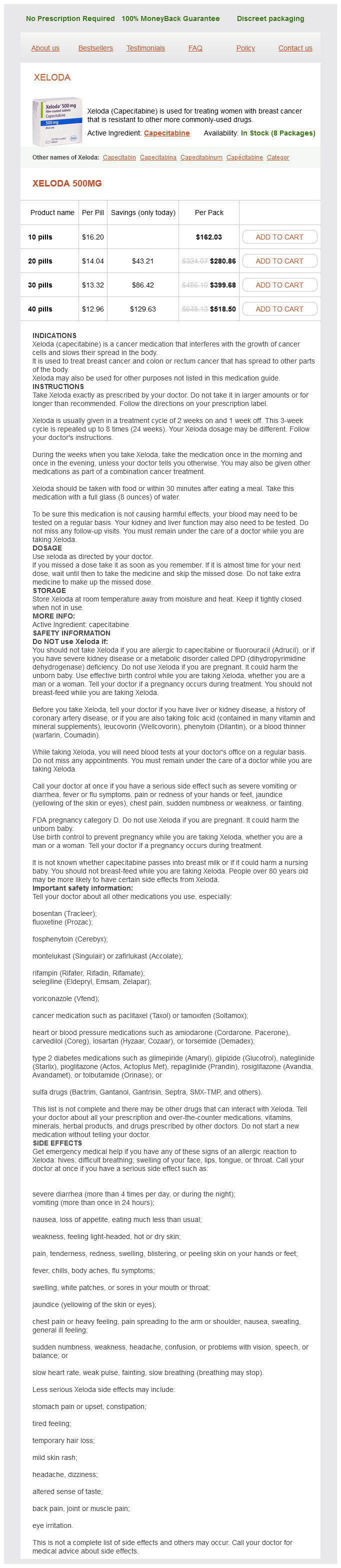
Buy capecitabine with paypal. Obstetrician Gynecologist: Nicole P. Kerner MD.
References
- Jones DT, Hutter B, Jager N, et al. Recurrent somatic alterations of FGFR1 and NTRK2 in pilocytic astrocytoma. Nat Genet 2013;45(8):927-932.
- Vacanti CJ, VanHouten RJ, Hill RC. A statistical analysis of the relationship of physical status to postoperative mortality in 68,388 cases. Anesth Analg 1970;49(4):564-566.
- Mariscal A, Balliu E, Diaz R, Casas JD, Gallart AM. Primary oat cell carcinoma of the breast: imaging features. AJR Am J Roentgenol 2004;183(4):1169-71.
- Daneman N, Gruneir A, Newman A, et al: Antibiotic use in long-term care facilities, J Antimicrob Chemother 66:2856n2863, 2011.
- Overgaard-Steensen C, Stodkilde-Jorgensen H, Larsson A, et al. Regional differences in osmotic behavior in brain during acute hyponatremia: an in vivo MRI-study of brain and skeletal muscle in pigs. Am J Physiol Regul Integr Comp Physiol. 2010;299:R521-R532.