Frank Craparo, MD
- Division Director
- Maternal Fetal Medicine
- Abington Memorial Hospital
- Abington, Pennsylvania
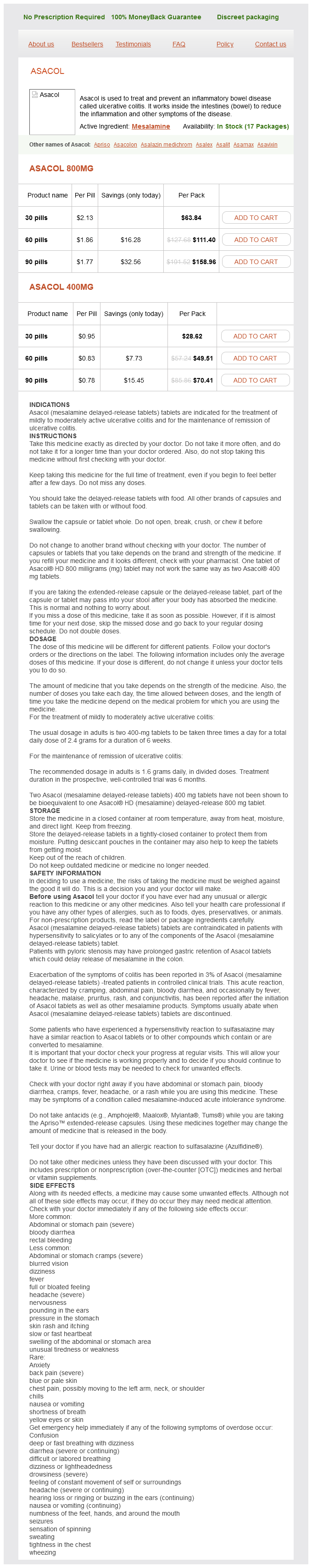
Pituitary stalk medications 2015 800 mg mesalamine mastercard, portal circulation in treatment 5th disease mesalamine 400 mg buy, 312 Pityriasis rosea treatment advocacy center 800 mg mesalamine purchase with visa, 887 Pityrosporum orbicularis medications not to be crushed generic 800 mg mesalamine mastercard, 882 Placenta abnormalities of medicine expiration dates order mesalamine with mastercard, 809-811 accreta, 809-811 infectious agents entering through, 191 Plaque dental, 473, 475 erythematous, in mycosis fungoides, 895 fibrous atheromatous, 320-321, 322/ complicated, 321, 323/ neuritic, 934 Plasma, 376 Plasma cells, 49, 57 in bone marrow, 377 chronic inflammation with, 209 neoplasms of, 461-467 perivascular, in skin infection, 216 Pseudobulbar palsy, 936 Pseudocyst, pancreatic, 676 Pseudogout, 983 Pseudohermaphroditism, 245 Pseudohypoparathyroidism, 86It, 862 Pseudolymphoma, 537 Pseudomembranous enterocolitis, C. Pyogenic granuloma, 477 Pyogenic liver abscess, 646 Pyogenic lymphadenitis, acute, 437 Pyogenic osteomyelitis, acute, 961-962 Pyogenic pericarditis, 370 Pyonephrosis, 722 Pyrexia, 169. Radiation cystitis, 741 Radiation dermatitis, 175/ Radiation myocarditis, 368 Radiation nephritis, 724 Radiation pneumonitis, 542 Radiation sterilization, 177 Radiation syndrome, acute, 173 Radicular cyst, 475 Radioisotope implants, 175 Radioisotopes, exposure to , cancer from, 286 Radiology. Renal adenocarcinoma, 731-734 clinical features of, 73If, 732-734 pathology of, 731-732, 733/ staging of, 734 Reproductive system, changes in, with aging, 248 Residual volume, 510 Resolution, 82 Respiratory acidosis, 25 Respiratory alkalosis, 25 aspirin and, 187 Respiratory disease. Respiratory obstruction, hypoxia from, 5 Respiratory syncytial virus acute bronchiolitis from, 218 Respiratory system, structure of, 506-507 Respiratory tract, upper, 484-491 infections of, 485 Reticulin, 27 Reticulocytes, 374 Reticuloendotheliosis, leukemic, 421 Retina, 492,497 Retinal detachment, 501 Retinal disease hypertensive, 327 vascular, 501 Retinitis pigmentosa, 500, 50 If Retinoblastoma, 289-290, 502-503 Retropharyngeal space, infections of, 485 Rubella, 882 congenital defects from, 240 cardiac, 341-342 perinatal infection with, 243 Rubella syndrome, 240 Rubor, 37 Rugal hypertrophy, 577 botryoides, 796, 958 high-grade, 990 low-grade, 990-991 Sarcoptes scabiei, 883 Satellite lesions, 893 Scabies, 883 Scarlet fever, erythrogenic toxin and, 205 Seborrheic keratosis, 889 Selenium, 162 Sella turcica, empty, 837 Seminoma, 752, 754/ Senile amyloidosis, 31 Senile atrophy, 252 Sensitization, 106 Sensitizing dose, 106 Septic infarcts, 129-130 Septic shock, 133 in acute pyelonephritis, 722 Septicemia, 195 Serologic tests for cancer diagnosis or follow-up, 308 Serosa, intestinal, 585 Serosal cavities, edema of, 24 Serotonin, in acute inflammation, 42-44 Serous cystadenocarcinoma, 770/, 771 Serous cystadenofibroma, 771 Serous cystadenoma of ovary, 770-771 of pancreas, 676 Serous inflammation, 45 Serous metaplasia, 254 Serous tumors, ovarian, 771 of low malignant potential, 770/, 771 Sertoli cell tumors, 755 Sertoli-Leydig cell tumors, 776 Serum, 376 Serum-sickness type reaction, 113 Sex, incidence of cancer and, 268-269 Sex chromosomes, 227 abnormalities of, 231-232, 245 Saphenous system, varicose veins of, 329/ Sarcoid myocarditis, 368 Sarcoidosis, 442, 542-543 clinical features of, 543, 861 Smoke inhalation, 167 Smoking, 181-183 acute erosive gastropathy and, 574 atherosclerosis and, 316 cancer and, 284 carcinoma of nasopharynx and, 486 chronic bronchitis and, 532 emphysema and, 532-533 esophageal cancer and, 568-569 fetal effects of, 241 lung cancer and, 547 pancreatic cancer and, 677 pharmacology and pathology of, 183 thromboangiitis obliterans and, 332 Social factors, incidence of cancer and, 270-271 Sodium, reabsorption of, in kidney, 693 Soft tissue neoplasms, 989-991 pathology of, 990-991 treatment of, 991 Solid infarcts, 129 Somatostatin, excess of, 689 Somatotropin. Sprue nontropical, 590 tropical, 591 Sputum examination of, in lung assessment, 507 infected, in secondary tuberculosis, 524 production of, in respiratory disease, 511 Squamous carcinoma of bladder, 745 of cervix, 790-794 of conjunctiva, 496 of eyelid, 494 laryngeal, 489^491 of lung, 548-549 of oral cavity, 477-478 of skin, 890 of vagina, 795-796 of vulva, 797 Squamous metaplasia, 254 of conjunctiva, 495-496 of endocervical epithelium, 256/, 790 Squamous papilloma, laryngeal, 489 Stable cells, 84 Staghorn calculus, 740/ Staphylococcus aureus acute bartholinitis from, 796 acute mastitis from, 818 cerebral abscess from, 219 Stress, acute erosive gastropathy and, 574 Stroke, 924-931 classification and etiology of, 926 Synovial sarcoma, 984 Synovitis, pigmented villonodular, 983 Syphilis, 801-805 aortic regurgitation in, 357 cardiovascular, 803-804 congenital, 243, 804-805 involvement of mouth in, 476 involvement of testis in, 750 latent, 803 neurologic involvement in, 803, 920, 921 Syringomyelia, 909 Systemic circulation, 312 Systemic venous congestion, 127 in heart failure, 340 T3. Testicular disease, manifestations of, 747-748 Testicular dysfunction, assessment of, 748-749 Testicular dysgenesis. Thyroglobulin, 855 Thyroglossal duct cyst, 838 Thyroid carcinoma, 850-855 anaplastic, 852 Tongue, inflammation of, riboflavin deficiency and, 160 Tonsillitis, acute, 484-485 Tonsils, infections of, 485 Tooth pulp abscess of, 475 infection of, 414-415 Tooth-forming tissues, neoplasms of, 475 Tophaceous gout, chronic, 982-983 Total lung capacity, 510 Totipotent cells, 264t neoplasms of, 264, 266/ Toxic agents, exogenous, cellular degeneration/necrosis from, 8 Toxic gases, pulmonary fibrosis from, 542 Toxic granulation, 47 Toxicity dose-related, 651 idiosyncratic, 651-652 Toxins, remotely acting, production of, by extracellular organisms, 204-205 Toxocara canis, 498 Toxoplasma gondii, 213 Transient ischemic attacks, 928-929 atheromatous embolism and, 148 Transitional cell carcinoma of bladder, 743-744. Trench foot, 165 Trench mouth, 476 Trephine biopsy, 376 Treponema carateum, pinta from, 881 pallidum meningitis from, 219 Trichomonas vaginalis acute vaginitis from, 794, 801 cervicitis from, 790 urethritis from, 801 Trichophyton, 882 Trichuris trichuria, 609 Tricuspid regurgitation, 358 Tricuspid stenosis, 358 Tricuspid valve, lesions of, 358 Triglycerides absorption of, in small intestine, 588-589 accumulation of, 8-10 intestinal, failure of removal of, malabsorption and, 590 normal metabolism of, in liver, 8, 9/ total plasma, atherosclerosis and, 316 Triiodothyronine (T3), 839-840. Ureaplasma urealyticum acute vaginitis from, 801 urethritis from, 801 Uremia, 695-696 platelet dysfunction from, 428 Ultrasonography, of thyroid nodule, 849 Ultrasound, effects of, 177 Ultraviolet radiation cancer from, 286 effects of, 176-177 Uncinate gyrus, herniation of, increased intracranial pressure and, 904 Undersea diving, pressure injury from, 165 Undifferentiated carcinoma of lung, 549-550 of ovary, 773 Unicameral bone cyst, 966 Urachal abnormalities, 740 Urate deposition, effects of, 982-983 Urate nephropathy, 725, 982-983 Vagina, 794-796 effects of aging on, 247 Fortunately, there are alternatives to the above drugs that do not readily cross the bloodbrain barrier (second-generation H1 antihistamines; doxycycline, tetracycline). Included are drugs with doses calculated per kilogram of body weight (isotretinoin, etretinate) and dose calculated per meter squared (bexarotene-Targretin). The question arises as to what to do with dosage calculations for very obese patients. Aside from treatment of panniculitis, there are virtually no indications for which the site of desired pharmacologic effect is in fatty tissue. Highly lipid-soluble drugs are readily distributed to fatty tissues, but when a steady state is reached, there is steady release back into the circulation. The bound drug is pharmacologically inactive, whereas the unbound drug = free drug = pharmacologically active drug. Acidic drugs are most commonly bound to albumin, whereas basic drugs bind preferentially to -1 acidic glycoprotein. There is a large circulatory reservoir for highly protein-bound drugs such as methotrexate. The second drug reservoir of interest is in various fatty tissues (including, but not limited to , subcutaneous fat) for highly lipophilic drugs, as discussed in the preceding paragraph. The third drug reservoir (the stratum corneum) pertains just to percutaneous absorption for topically applied medications. In all three settings the free drug and the drug in the reservoir are in equilibrium. As the free drug is metabolized and excreted, corresponding amounts of the drug in these tissue and circulatory reservoirs are released into the free/ active drug fraction. Pharmacodynamics (The Drug Produces the Desired Pharmacologic Effect) the subject of pharmacodynamics is very complicated. Considering all the diverse mechanisms of actions discussed in this book (let alone the diversity of drug mechanisms in the entirety of medicine), it is not possible to summarize general principles behind all of them. In contrast, it is possible to cover a few areas of central importance to understanding pharmacodynamics. These include the concepts of drug receptors, enzyme inhibition by drugs, signal transduction, and transcription factors. A systemic drug with a relatively low bioavailability is acyclovir; the prodrug for acyclovir, valacyclovir, has at least three times greater bioavailability. At the other end of the spectrum are the fluoroquinolones, for which oral absorption (and resultant bioavailability) is so complete that the oral and intravenous doses for many members of this drug group are identical. A more optimal method (if it were more practical) would be to calculate bioavailability at the site of intended action; for drugs discussed in this book, it would be based on tissue levels at the site of intended action, the various skin structures. For most chapters in this book that discuss systemic drugs there are tables that present data for the following: (1) % bioavailable and (2) % protein binding. Changes in albumin levels in disease states such as severe liver or renal disease will often necessitate drug dosage adjustments for drugs (such as methotrexate) that are highly protein bound. Creating drug formulations with a more optimal bioavailability is a daunting task for the pharmaceutical industry. In the past few decades there have been updated formulations of older drugs with higher bioavailability, more predictable bioavailability, or both. For drugs with a relatively narrow therapeutic index (cyclosporine, methoxsalen), improved predictability of the drug absorption and resultant bioavailability are very important. The release of Neoral and Gengraf (in place of the previous cyclosporine formulation, Sandimmune) is an example for both improved % bioavailability and more predictable bioavailability of the newer formulation. Likewise, Oxsoralen Ultra demonstrates improvement in both of these two parameters. In a separate example, the need for improved efficacy from griseofulvin led to the progression from the original griseofulvin formulations microsize formulations ultramicrosize formulations. Each step of this progression resulted in improved bioavailability and smaller griseofulvin dosages required for an adequate therapeutic response. Reduce an undesirable physiologic or pharmacologic response (drug antagonists or receptor blockers); or 5. Only a proportion of these concepts can be realistically addressed in the remainder of this section on pharmacodynamics. In this definition, any molecule to which a drug binds, thus initiating an effector mechanism leading to a specific pharmacologic response, is a drug receptor. The drug receptor subtypes that are easiest to characterize are cell surface receptors for endogenous neurohormonal ligands. In addition, lipophilic drugs easily absorbed through cellular membranes may have cytosolic drug receptors. A few examples of drugs that are either antagonists or agonists at well-defined cellular receptors are given in Table 1. A number of drugs representing antimicrobial agents for bacterial, viral, and fungal infections capitalize on vital enzyme systems, which are more readily inhibited in the infectious organism than in the human host. Finally, a number of drugs inhibit enzyme systems that contribute important downstream mediators to an inflammatory response. Drugs that inhibit enzyme systems of importance to Signal Transduction and Transcription Factors these two aspects of pharmacodynamics have a number of conceptual similarities, albeit with very distinctive mechanisms of action. Tremendous details on the various receptor/signal transduction categories (six main families) are beyond the scope of this chapter but are available in the Bibliography. Calcium is an important component of the above T-cell signal transduction system in two locations; calcineurin is a calcium-dependent enzyme, with a calcium-binding protein (calmodulin) playing an important role as well. The polyamine pathway creates a process known as inflammatory hyperplasia, which is an important component of the pathogenesis of both psoriasis and various malignancies. Retinoids inhibit the activity of ornithine decarboxylase, the ratelimiting enzyme in the polyamine pathway. This signal transduction enzyme inhibition is important to the benefits of systemic retinoids in both psoriasis therapy and retinoid chemoprevention of cutaneous malignancies in solid organ transplantation patients. This transcription factor is pivotal in the upregulation of a multitude of cytokines of central importance in the inflammatory response to a wide variety of stimuli. The result of these enzymes is a somewhat more hydrophilic (water-soluble) metabolite, which may provide a site of attachment for subsequent conjugation reactions. An example of this principle would be the itraconazole metabolite hydroxyitraconazole, which also has significant antifungal activity. The topic of pharmacogenetics largely addresses genetically based variations in the above metabolic enzyme systems. Most drugs are metabolized by phase I (oxidation Toxicity/toxic effect aConfusing reality is that immunologic hypersensitivity may occur due to excessive quantities of a reactive metabolite, rendering immunogenic a previously normal endogenous protein (see Chapter 62 Hepatotoxicity of Dermatologic Drug Therapy). The excreted drug can be the parent drug, drug metabolites, or combinations of both. An example would be fluconazole, which because of its relatively hydrophilic properties has a significant portion of the administered drug excreted through the kidney unchanged. In particular, greater hydrophilicity favors renal excretion, which has a much larger overall capacity for drug excretion than the hepatobiliary route. In reality, the drugs discussed in this book are frequently excreted by several of the above routes, both as free drug and as a variety of metabolites. The reader should also be aware that many drugs conjugated in the liver, and excreted into bile, will subsequently undergo hydrolysis in the small intestine and be reabsorbed (enterohepatic recirculation) through many cycles. It is very important to recognize that disease-induced or agedependent reduction in renal function should prompt the clinician to significantly reduce dosages of drugs with significant renal clearance. An example would be the increased risk for pancytopenia and other complications with methotrexate when standard doses are administered to patients with either disease- or agerelated reduction in renal function. Likewise, drugs that have significant liver metabolism and excretion should have dosage reductions with advanced liver disease. The conversion of prednisone (prodrug) to prednisolone (active form) is dependent on a hepatic-based enzyme, which in end-stage liver disease may not produce therapeutically adequate quantities of the active drug form prednisolone. Once the prodrug is metabolized to the active drug, the principles of interest follow through the distribution, bioavailability, and pharmacodynamics sections as with other drugs already in active form once absorbed. Percutaneous Absorption General Principles There is a wealth of scientific and practical information in Tables 1. Drug diffusion coefficient-the ability of the drug (due to innate molecular properties) to penetrate through all layers of skin once in the stratum corneum; 4. Drug concentration-the specific drug concentration of a given topical product; and, 5. Superficial dermal vascular plexus-site of systemic absorption for topically applied drugs. It is important to note here that all topical drug absorption occurs via passive diffusion. Topical medications applied in several clinical settings can produce immediate hypersensitivity (Coombs-Gell type I) reactions. In particular, topical application to ulcerated skin can give the applied medication almost immediate access to systemic circulation. There have been reports of anaphylaxis to topical bacitracin or neomycin in this setting. Although the risk from topical application of medications to these above sites is usually small, the clinician should always be mindful of this systemic absorption potential. In general, the choice of vehicle is just as important as choosing the proper active ingredient. The good news is that this is an easily reversible process, particularly if the clinician is mindful of the potential for tachyphylaxis. Transdermal Medication Formulations One final routine of topical administration of medications that is tangentially related to dermatology deserves mention here. The potential for certain drugs to have reduced bioavailability through excessive hepatic and small intestine first-pass metabolism can be circumvented by transdermal administration. An excellent example would be transdermal estrogen administration, which allows the drug to be absorbed directly into systemic circulation.
The basic defect is an impermeability of epithelial cell membrane to chloride ions medicine park cabins cheap mesalamine 400 mg online. The cystic fibrosis allele codes for a membrane-associated protein that serves as a chloride channel symptoms and diagnosis generic mesalamine 800 mg with visa. This defect results in (1) increased salt content of sweat and (2) decreased water content of respiratory aquapel glass treatment purchase mesalamine 800 mg overnight delivery, intestinal medicine rash discount 800 mg mesalamine with visa, and pancreatic exocrine secretion treatment lead poisoning discount mesalamine 800 mg without prescription, leading to increased viscosity. Pancreatic ducts are plugged with mucus, leading to chronic inflammation, with atrophy of acini, fibrosis, and dilation of ducts (hence the term fibrocystic disease). Lack of pancreatic lipase in the intestine causes maldigestion of fat, leading to steatorrhea. Pulmonary changes (see Chapter 35) are the most serious manifestation of cystic fibrosis. Bronchial mucus plugs lead to collapse of the lung, recurrent infections-including lung abscess-and ultimately fibrosis and bronchiectasis. Increased amounts of viscid intestinal mucus in the lumen may result in intestinal obstruction in the neonatal period (meconium ileus). Bile duct obstruction may result in jaundice; changes in the vas deferens and seminal vesicles may cause infertility in the male. A sweat sodium level in excess of 60 meq/L is diagnostic in a patient with clinical features of cystic fibrosis. Twenty years ago, survival beyond infancy was unusual; today, most individuals survive to adulthood, although life expectancy is still much less than normal. Because ectopic pancreatic tissue is a developmental mass composed of disorganized pancreatic acini, ducts, and muscle that do not belong in these locations, the term choristoma may be used. The most important is a rare malformation of the head of the pancreas, which results in a complete collar of pancreatic tissue around the second part of the duodenum (annular pancreas); this may result in duodenal constriction and obstruction. Many of the features of this disease are caused by obstruction of exocrine ducts due to the increased viscosity of secretions. It is associated with extensive destruction of pancreatic and peripancreatic tissue and acute inflammation. Acute pancreatitis is a common and important medical emergency, accounting for about one in every 500 admissions to general hospital emergency rooms. Etiology In about 25% of cases of acute pancreatitis, no etiologic factor can be identified. Infectious agents are usually not involved, although mild nonnecrotizing acute pancreatitis occurs in association with some viral diseases-commonly mumps and cytomegalovirus infection. Biliary Tract Calculi: Biliary tract calculi are present in about 50% of cases and may obstruct the terminal bile duct. Reflux of bile or infected duodenal contents into the pancreatic duct has been suggested as a mechanism leading to pancreatitis in bile duct disease. Obstruction of the pancreatic duct may occur when a calculus becomes lodged in the ampulla of Vater. Acute pancreatitis complicating gallstones is chiefly a disorder of women because of the female preponderance of gallstone disease. Alcoholism: Alcoholism as a cause of acute pancreatitis occurs with varying frequency in different parts of the world. It is common in the United States, being involved in 65% of cases of acute pancreatitis; in Europe, the incidence is 5-20%. Hypercalcemia: Hypercalcemia, as occurs in primary hyperparathyroidism, is complicated by acute pancreatitis in about 10% of cases. A high plasma calcium concentration is thought to stimulate activation of trypsinogen in the pancreatic duct. Hyperlipidemias: the hyperlipidemias-particularly those types associated with increased plasma levels of chylomicrons-are complicated by acute pancreatitis. It is postulated that free fatty acids liberated by the action of pancreatic lipase produce acinar injury. Acute pancreatitis-etiologic factors, mechanisms, pathologic changes, and clinical effects. Drugs and Radiation: Thiazide diuretics, corticosteroids, anticancer agents, and other drugs may also cause acute pancreatitis. Radiation to the retroperitoneum for treatment of malignant neoplasms is an uncommon cause of acute pancreatitis. Trypsin and chymotrypsin activate phospholipase and elastase as well as kinins, complement, the coagulation cascade, and plasmin, leading to acute inflammation, thrombosis, and hemorrhage. Pancreatic lipase acts on surrounding adipose tissue, causing enzymatic fat necrosis (see Chapter 1). Circulating amylase does not contribute to cell injury; however, phospholipases are thought to contribute to the production of adult respiratory distress syndrome by interfering with the normal function of pulmonary surfactant. Rarely, high serum lipase levels are associated with fat necrosis at sites distant from the pancreas. Pathology Acute pancreatitis is characterized by widespread necrosis in tissues subjected to the effect of extravasated pancreatic enzymes. Activation of the plasma coagulation cascade may lead to disseminated intravascular coagulation. Laboratory Studies There is an almost immediate (within hours) elevation of the serum amylase, often to 10-20 times the normal upper level; amylase levels return to normal in 2-3 days. Transient glycosuria is present in the acute stage in about 10% of cases as a result of islet dysfunction. Permanent diabetes mellitus almost never follows a single attack of acute pancreatitis. Complications Most patients recover from the acute attack with proper supportive care, and the pancreas regenerates and returns almost to normal, with mild residual scarring. In severe cases, death may occur in the acute phase as a consequence of pancreatic abscess, severe hemorrhage, shock, disseminated intravascular coagulation, or respiratory distress syndrome. Pancreatic pseudocyst (see below) may follow weeks to months after recovery from an acute attack. Acute pancreatitis, showing marked hemorrhagic necrosis in the upper retroperitoneum around the pancreas. In severe cases, massive liquefactive necrosis of the pancreas occurs, resulting in a pancreatic abscess. In very severe cases, death may occur before an adequate inflammatory response can be mobilized. The peritoneal cavity sometimes contains a brownish serous fluid (pancreatic ascites). This fluid contains altered blood, fat globules ("chicken broth"), and very high levels of amylase. Patients develop severe constant epigastric pain, frequently referred to the back, accompanied by vomiting and shock. Etiology Chronic alcoholism and biliary tract calculi are the two main conditions that are associated with chronic pancreatitis. In the United States, alcoholism is believed to be implicated in about 40% of cases and biliary tract disease in 20%. Some cases follow recurrent acute episodes; cystic fibrosis is a specific type of chronic pancreatitis described earlier. The changes usually involve the gland diffusely; more rarely, a firm, localized mass forms that is difficult to distinguish grossly from carcinoma. The pancreatic ducts show multiple areas of stenosis with irregular dilation distally. Chronic pancreatitis-etiologic factors, pathologic changes, and clinical effects. At the end stage there are almost no recognizable acini, and the gland is composed of dilated ducts separated by collagen. It may be constant or intermittent and can be so severe as to lead to narcotic dependence. In many cases, pain is associated with acute exacerbations, and the patients are asymptomatic between relapses. When pain is caused by dilation of the duct system, surgical correction by draining the dilated duct system may provide relief. Pancreatic exocrine insufficiency due to failure of secretion of pancreatic juice leads to steatorrhea, malabsorption of fat-soluble vitamins, and weight loss. Many patients have recurrent attacks of severe pain, vomiting, and elevation of serum amylase, due probably to repeated acute episodes (chronic relapsing pancreatitis). The pancreatic parenchyma between the ducts has undergone marked atrophy and fibrous contraction. This condition is difficult to differentiate from jaundice due to pancreatic carcinoma. There are no specific laboratory tests, but the presence of calcification on x-ray provides supportive evidence. In chronic disease, the amount of residual pancreatic tissue may be insufficient to cause elevation of serum amylase. Treatment & Prognosis Treatment of chronic pancreatitis consists of management of the pain, malabsorption, and diabetes. When pain cannot be controlled by drugs, surgery to drain the pancreatic duct (by creating an opening between the duct and a loop of jejunum, ie, pancreaticojejunostomy) often has good results. Malabsorption and diabetes mellitus can be controlled by dietary supplements and insulin if necessary. Treatment consists of establishing surgical drainage of the cyst either into the stomach or into a loop of jejunum. Complications of pancreatic pseudocyst include (1) acute rupture of the cyst into the intestine, most commonly the stomach and transverse colon, producing intestinal hemorrhage, which may be severe; (2) secondary infection, leading to the formation of an abscess; and (3) compression of the common bile duct, causing obstructive jaundice. Congenital pancreatic cysts are often associated with polycystic renal disease and congenital hepatic fibrosis. Congenital pancreatic cysts also occur in von Hippel-Lindau disease (see Chapter 62). A pseudocyst is usually a solitary fluid-filled unilocular structure of variable size lined by a wall composed of collagen and inflamed granulation tissue. Pseudocysts usually occur after an attack of acute necrotizing pancreatitis or during chronic relapsing pancreatitis and probably represent the end result of hemorrhagic necrosis, with the liquefied material walled off by granulation tissue and fibrosis. They contain brownish serous fluid composed of pancreatic juice with a high enzyme content and altered blood. Most pseudocysts occur in and around the pancreas; rarely, when pancreatic enzyme leakage produces necrosis away from the pancreas, pseudocysts may occur at a considerable distance from the pancreas (eg, in the right iliac fossa). Patients with pseudocysts present with an abdominal mass, and most give a history of abdominal pain Serous cystadenoma is a rare solitary, benign cystic neoplasm of the pancreas. It may reach large size but rarely causes symptoms, and it is most commonly an incidental finding at abdominal surgery or radiography. It consists of multiple small locules lined by cuboidal epithelium, the cells of which contain abundant glycogen (also called microcystic, serous, and glycogen-rich cystadenoma). The cysts are lined by tall columnar epithelium and contain a glairy mucinous fluid. The malignant counterpart shows cytologic atypia, stratification of cells, and invasion of the capsule. Mucinous cystadenocarcinoma behaves like a slowly growing low-grade malignant neoplasm. Incidence & Etiology Approximately 25,000 patients annually in the United States die from pancreatic carcinoma, accounting for 6% of cancer deaths; the incidence is slightly higher in men than in women and is increasing. A large number of dietary factors have been proposed, including decaffeinated coffee and high-fat diets. Carcinomas of the head tend to obstruct the common bile duct early in their course and present at a stage when the tumor is small. Pancreatic carcinoma frequently evokes marked fibrosis; it may distort the duodenal loop, producing a typical "inverted 3" appearance on barium x-ray studies. Microscopically, over 90% of cases are well-differentiated adenocarcinomas, associated with marked fibrosis. Contrast the normal ductal epithelial cells on the left with the greatly enlarged and pleomorphic carcinoma cells on the right and in the lumen. Well-differentiated carcinoma of the pancreas, showing perineural invasion (the nerve is at the bottom center of this photograph). Rarely, acinar cell carcinomas secrete lipase into the bloodstream and cause fat necrosis in the subcutaneous tissue and bone marrow throughout the body. Bloodstream spread also occurs early, with the liver being the most common site of secondary deposits. Clinical Features Carcinoma of the head of the pancreas presents with common bile duct obstruction. A high proportion of patients present with evidence of metastatic disease, most often in the liver. Skin rashes and lytic bone lesions due to fat necrosis may be present in lipasesecreting acinar cell carcinomas. Carcinoembryonic antigen levels in the serum are elevated in some cases; this is not a specific finding, as colon, lung, and other cancers may also show elevated levels. The presence of a more specific pancreatic oncofetal antigen has been reported recently in a high proportion of cases, but data are preliminary. Computerized tomography is effective in establishing the presence of a solid mass. Percutaneous fine-needle aspiration of the mass under radiologic guidance provides tissue for cytologic examination and is an excellent method of making the diagnosis. Rarely, patients with pancreatic carcinoma develop disseminated intravascular coagulation, due probably to thromboplastic substances present in the mucinous product of the adenocarcinoma.
Incidence the incidence of anemia varies from 2% to 15% in different studies in Britain and the United States internal medicine discount mesalamine 800 mg free shipping. Mild anemia without symptoms is quite common medicinenetcom purchase 400 mg mesalamine amex, but anemia may be the presenting sign of serious disease 98941 treatment code 800 mg mesalamine order with visa. If normal range American values are used medicine evolution buy discount mesalamine line, anemia is much more common in underdeveloped countries; this observation raises questions about what the normal range really is and whether it should be defined differently for different populations medicine lookup 400 mg mesalamine purchase fast delivery. Diagnosis When a patient presents with symptoms of anemia-eg, pallor of mucous membranes, fatigue, and breathlessness-the initial diagnosis may be confirmed by demonstration of a decreased hemoglobin level. Identification of the cause of anemia may require bone marrow examination, hemoglobin electrophoresis, and determination of serum iron, vitamin B12, and folate levels. The tests required will vary with different patients, eg, bone marrow examination is critical in macrocytic anemia and of limited value in microcytic-hypochromic anemia. Because there is no mechanism to decrease the rate of peripheral destruction of erythrocytes, anemia results. Etiology Drugs are the most common cause of aplastic anemia in clinical practice (Table 24-4). Pathology & Clinical Features Aplastic anemia shows a markedly hypocellular bone marrow with a reduction of all cell lines. Predictable, dose-related: Chiefly cytotoxic anticancer drugs (methotrexate, mechlorethamine, vincristine, daunorubicin, etc) b. Unpredictable idiosyncratic reactions: Rare; do not require prior exposure, nonimmune Antiepileptic drugs (hydantoins) Oral antidiabetic agents (tolbutamide, chlorpropamide) Tranquilizers (chlorpromazine, chlordiazepoxide, etc) Antirheumatic drugs (phenylbutazone indomethacin, colchicine, gold salts) Antibacterial agents (sulfonamides, streptomycin, isoniazid, tetracyclines, choramphenicol2) Many others (hydralazine, quinacrine, thiazide diuretics) c. Unpredictable hypersensitivity: Immune reaction; requires prior exposure; many drugs (antibodies to drugs often detectable) 2. Radiation: Either external beam or radioactive isotopes Associated diseases Familial hypoplastic anemia (Fanconi; chromosomal abnormalities and a high incidence of leukemia) With infective hepatitis With pancreatitis With paroxysmal nocturnal hemoglobinuria Preleukemia 1 Note: May present with total marrow failure, failure of red cell production alone or failure of white cell production alone, (neutropenia). This disorder frequently remits spontaneously or in response to corticosteroid therapy. Acquired red cell aplasia is usually a transient complication that occurs in congenital hemolytic anemias such as sickle cell anemia. Adult red cell aplasia may also occur as a complication of thymoma by an unknown mechanism. Replacement of proliferating marrow cells of sufficient degree may result in anemia, leukopenia, or thrombocytopenia. Compensatory phenomena include (1) the development of active marrow in sites outside the marrow cavity (extramedullary hematopoiesis or myeloid metaplasia, especially in spleen and liver) and (2) release of cells from the marrow before maturation is complete. The latter results in a shift to the left (immature white cells, including myeloblasts and myelocytes) and the presence of nucleated red cells (normoblasts) in the peripheral blood-hence the designation leukoerythroblastic anemia. Granulocytopenia: Granulocytopenia (neutropenia) occurs rapidly because these cells have a very short life span; infections and oral ulcers result. In severe, irreversible cases, death results from the effects of infections (eg, pneumonia, septicemia), bleeding (eg, intracranial hemorrhage), or anemia (eg, cardiac failure). Megaloblastic anemias are a subset of macrocytic anemias in which the maturation phase of erythropoiesis in the bone marrow is abnormal, resulting in erythroid precursors that are enlarged and show failure of nuclear maturation (megaloblasts). Etiology Megaloblastic anemias result from conditions in which nucleic acid synthesis is abnormal, as in vitamin B12 and folic acid deficiencies (Table 24-5). Note that vitamin B12 reservoirs in the liver are normally sufficient for several years; following gastrectomy (which removes the source of intrinsic factor, thereby reducing vitamin B12 absorption), a decade may pass before vitamin B12 megaloblastic anemia becomes apparent. Vitamin B12 is present in high concentration in animal liver and to some degree in most meats but is absent in plants. The late megaloblast, for example, shows a primitive nucleus and fully hemoglobinized cytoplasm-in contrast with the late normoblast, which has a pyknotic nucleus. The bone marrow is hypercellular and contains large numbers of early megaloblasts; prior to the discovery of the beneficial effect of liver extract (contains vitamin B12) on pernicious anemia, this picture was mistaken for that of acute leukemia. Oval forms (macro-ovalocytes) are prominent, and Ho well-Jolly bodies, consisting of nuclear debris, are occasionally seen. Megaloblastic anemias are therefore macrocytic anemias if morphologic classification is used. Lack of vitamin B12 leads to abnormal myelin synthesis, possibly via a deficiency of methionine (B). Neutrophil precursors in the bone marrow show marked enlargement; giant metamyelocytes are characteristic. These include the intestinal mucosa and other epithelia, which show cell enlargement and nuclear abnormalities. Recognition of these changes is important contextually if cytologic studies are undertaken-eg, in uterine cervical smears, the nuclear changes of folate deficiency may resemble those of dysplasia. Clinical Features & Diagnosis Patients with megaloblastic anemia present with symptoms of severe anemia. Megaloblastic anemia should be suspected upon finding macrocytic anemia with hypersegmented neutrophils in the peripheral blood. Bone marrow examination is necessary for confirmation and shows megaloblastic erythropoiesis. Establishment of the precise cause of megaloblastic anemia requires further clinical examination and laboratory testing (Table 24-6). There are two principal forms of megaloblastic anemia: folate deficiency, which has several underlying causes (Table 24-5); and vitamin B12 deficiency, again with several causes, including pernicious anemia, which will be discussed later in this chapter at some length. Vitamin B12 Deficiency Megaloblastic anemia Peripheral blood features Subacute combined degeneration of the spinal cord Serum vitamin B121 Serum folate1 Red cell folate Response to vitamin B12 by injection2 Folate Deficiency + Identical + Normal + Low Normal Normal Low Low + Normal - Vitamin B12 absorption test Abnormal3 (Schilling) Antiparietal or intrinsic factor antibodies in serum 1 2 ±4 - Serum levels measured by radioimmunoassay. It is normal in dietary deficiency and abnormal in bacterial overgrowth, general malabsorption, and pernicious anemia; in pernicious anemia it corrects to normal if intrinsic factor is added. Note that the megaloblastic anemia of folate deficiency is identical to that of vitamin B12 deficiency; administration of folic acid may thus mask (partially correct) the anemia of vitamin B12 deficiency, and vice versa; however, folic acid administration does not correct the neurologic effects of B12 deficiency. For this reason, it is important to exclude vitamin B12 deficiency before treating megaloblastic anemia with folate. This process is associated with failure of secretion of acid and intrinsic factor. Achlorhydria is invariably present in these patients and can be demonstrated by sampling gastric fluid after appropriate stimulation tests. In the absence of intrinsic factor, vitamin B12 absorption is drastically reduced. Three types of autoantibodies may be demonstrated in both serum and gastric juice: (1) About 75% of patients have an antibody that blocks vitamin B12 binding to intrinsic factor (blocking antibody); (2) about 50% have an antibody that binds with the intrinsic factor-vitamin B12 complex, interfering with the binding of the complex to ileal mucosal receptors, a prerequisite for vitamin B12 absorption; and (3) about 90% of patients have antibodies against gastric parietal cells. While useful for the diagnosis of pernicious anemia, the role played by these autoantibodies in producing the disease is uncertain. A few patients with pernicious anemia have none of the three antibodies, and the disease does occur in patients with hypogammaglobulinemia. The demonstration of cell-mediated immunity (T cell hypersensitivity) against intrinsic factor is therefore of particular interest. Megaloblastic anemia and other changes resulting from failure of nucleic acid synthesis have been described above. The most characteristic neurologic abnormality in pernicious anemia is subacute combined degeneration of the spinal cord, which is characterized by demyelination of the posterior and lateral columns of the cord (Chapter 64). This leads to paresthesia, loss of position and vibration sense, weakness, ataxia, and finally spasticity. These neurologic changes do not occur in patients with folic acid deficiency (Table 24-6). Treatment & Prognosis Patients with pernicious anemia die without treatment (the adjective pernicious indicates the high Pernicious anemia is a form of megaloblastic anemia due to vitamin B12 deficiency. It is common (affecting up to 1% of adults over 60 years of age) in western Europe and among Caucasians (particularly of Northern European descent) in the United States; however, it is rare in Asia and Africa. Pernicious anemia occurs predominantly after the age of 50 years, slightly more often in males than in females. Family members show an increased incidence of achlorhydria, decreased vitamin B12 absorption, or overt pernicious anemia. Pathogenesis Adult pernicious anemia is an autoimmune disease, caused by immunologic destruction of the gastric mucosa. With adequate replacement therapy with injected vitamin B12 (cyanocobalamin), these patients live a normal life. Patients with pernicious anemia remain at increased risk for development of gastric carcinoma in the atrophic gastric mucosa and must be followed carefully for this complication. Precancerous epithelial dysplasia usually occurs before invasive carcinoma develops and can be detected in gastric biopsies. It is common in underdeveloped as well as developed countries, but the causes differ in the two settings. In the United States, pregnancy and chronic blood loss due to gastrointestinal ulcers or neoplasms are the most common causes. It has been estimated that in the United States, 50% of pregnant women, 20% of all women, 20% of preschool children, and 3% of men suffer from iron deficiency. Normal plasma has enough transferrin (iron binding capacity) to bind 250-400 jig of iron per deciliter of blood. Upon demand, absorption of iron can increase only to a limited degree (to about 20% of dietary iron). Dietary Deficiency: the daily dietary iron requirement is 5-10 mg/d (equivalent to 0. Possible causes Infancy and childhood Dietary Chronic blood loss Chronic infection Prematurity Adult female (reproductive years) Dietary Excessive menstrual loss Pregnancies and spontaneous abortions Gastrointestinal blood loss Hematuria, other blood loss Adult male (and postmenopausal female) Gastrointestinal blood loss Drugs (eg, aspirin) Dietary (rare) Epistaxis, hematuria, other blood loss Hereditary hemorrhagic telangiectasia1 Investigations History Diet Drugs (aspirin) Gastrointestinal symptoms (pain, indigestion, black or bloody stools, dysphagia) Menstrual history Examination Rectal, sigmoidoscopy (cancer, hemorrhoids) Pelvic, in female Evidence of telangiectases X-rays Barium swallow: esophageal varices Barium meal: peptic ulcer, gastric cancer, hiatus hernia Barium enema: carcinoma, colitis Chest: lung cancer, other causes of hemoptysis Fecal occult blood2 Tests for blood in feces Urinalysis3 Tests for blood in urine 1 Osler-Weber-Rendu disease: Hereditary hemorrhagic telangiectasia produces multiple pinpoint bleeding foci in the gastrointestinal tract, mouth, lips, and skin. Chronic Blood Loss: Loss of blood is a major cause of iron deficiency throughout the world. One hundred milliliters of whole blood with a hemoglobin of 15 g contains about 50 mg of iron (1 g hemoglobin contains 3. Excessive menstrual blood loss from any cause and occult gastrointestinal blood loss (due to hookworm infection, peptic ulcer disease, chronic aspirin ingestion, esophageal varices, hemorrhoids, and neoplasms) are the most common causes of iron deficiency anemia; chronic loss of as little as one teaspoonful (5 mL) of blood per day will tip most people into negative iron balance. However, with chronic loss the onset of iron deficiency interferes with erythropoiesis, and this leads to anemia. Pathology Iron deficiency is first reflected in a decrease in iron stores, demonstrated by a decreased serum ferritin level and an absence of iron in a specimen of bone marrow. The main effect of iron deficiency is to reduce erythropoiesis, with several consequences. Anemia: Anemia is due both to a decreased amount of hemoglobin in individual red cells and to a decrease in the total red cell mass. Hypochromic microcytic anemia is the typical result of iron deficiency; however, it is not specific for iron deficiency anemia. The normoblasts are small, have decreased hemoglobin, and show an irregular frayed cell membrane. Storage iron is absent, and the number of sideroblasts (normoblasts containing cytoplasmic iron) is decreased (ie, the Prussian blue stain for iron is negative). Epithelial Changes: Iron deficiency also results in atrophy of many epithelial surfaces, eg, the 15 mg of iron, marginally adequate for women. Iron deficiency due to dietary deficiency occurs commonly in underdeveloped countries. Increased Demand for Iron: In developed countries, iron deficiency occurs in individuals on marginal diets or when there is an increased demand for iron, ie, in the growth phase of early childhood and during pregnancy and lactation. Malabsorption of Iron: Iron deficiency may occur in severe generalized malabsorptive states such as celiac disease and tropical sprue. Koilonychia (concave fingernails with abnormal ridging and splitting) is a specific result of iron deficiency. The association of iron deficiency anemia, koilonychia, atrophic glossitis, and dysphagia is known as Plummer-Vinson syndrome (Chapter 37). Treatment It is vital to establish the underlying cause prior to treatment (Table 24-7). Iron replacement, either orally or parenterally, is effective in treating iron deficiency anemia and replenishing body stores. It is important in the management of patients with iron deficiency anemia to find and correct the reason for anemia. The anemia in these cases is caused by failure of transport of storage iron into the plasma and therefore into developing erythrocytes. Iron therapy does not help and may actually cause harm by increasing the tissue iron stores. Sideroblastic anemias are characterized by either a hypochromic, microcytic, or dimorphic peripheral blood erythrocyte appearance. A dimorphic peripheral blood picture is one in which there is an admixture of hypochromic, microcytic erythrocytes and macrocytic erythrocytes. The marrow shows erythroid hyperplasia, markedly increased amounts of storage iron, increased number of sideroblasts, and defective hemoglobinization. A characteristic feature is the presence of ring sideroblasts-normoblasts in which the amount of iron is so greatly increased that it appears as cytoplasmic granules arranged in a complete ring around the nucleus. It appears that there is a defect in incorporating iron into the hemoglobin molecule within the erythrocyte. In general, the number of sideroblasts is greater in primary acquired Sideroblastic anemia than in the secondary type of disease. In some patients with refractory Sideroblastic anemia, increased numbers of immature white cells (mainly myeloblasts) are present. This is believed to be a premalignant change (dysmyelopoiesis), with many (10%) of these patients going on to develop acute leukemia. In the acute bleeding phase, blood values-including red cell count, hemoglobin, and hematocrit-are normal because equivalent amounts of red cells, hemoglobin, and plasma are lost. An important compensation for hypovolemia is retention of water and electrolytes by the kidneys to restore blood volume. Within hours, the effect of this dilution is seen as a progressive decrease in red cell count, hemoglo- Patients with chronic renal failure develop a normochromic, normocytic anemia due to failure of normal erythropoietin secretion by the kidney.
The stage may be the clinical stage symptoms 7dp5dt cheap mesalamine 400 mg mastercard, which is determined by the history and physical examination symptoms vertigo purchase generic mesalamine online, radiologic studies medications that cause hair loss cheap mesalamine 400 mg online, isotopic scans treatment plant cheap mesalamine 800 mg online, laboratory tests of urine treatment 3rd degree heart block order cheapest mesalamine and mesalamine, blood, and liver, and the initial biopsy results; or the pathologic stage, which is based on histologic findings in tissue removed by biopsy or laparotomy, with symbols indicating the tissue samples taken (N = node, H = liver, S = spleen, L = lung, M = marrow, P = pleura, O = bone, D = skin) and the results of histopathologic examination (+ indicates involved, indicates not involved). It is useful to classify malignant lymphomas as low-grade, intermediate-grade, and high-grade because the grades correlate well with survival. In general, lymphomas with a follicular histologic pattern are of lower grade (longer survival times) than those with a diffuse pattern. The neoplastic cells resemble B immune-blasts, with large nuclei, prominent nucleoli, abundant basophilic cytoplasm, and a high mitotic rate. Mycosis fungoides (cutaneous T cell lymphoma), showing infiltration of dermis and epidermis by T cells characterized by irregular nuclei with lobation (cerebriform nuclei). The dotted line represents the plane of the epidermal basement membrane, which has become obliterated. It must be emphasized that the clinical and pathologic staging classifications are unique to the main subtypes of lymphomas and apply only at the time of disease presentation-before definitive therapy is started. The use of combined (multiple-agent) chemotherapy (Table 29-10) has favorably influenced the, prognosis of these neoplasms. Paradoxically, treatment of previously poor prognostic types of lymphoma (lymphoblastic and immunoblastic) is sometimes more successful in providing complete remission than treatment of the less aggressive histologic types. This probably reflects the fact that most chemotherapeutic agents act only on dividing cells, which are plentiful in high-grade lymphomas but sparse in low-grade lymphomas. Cyclic chemotherapeutic regimens are used so that neoplastic cells that are not in the dividing phase during one dose and therefore survive are treated by the next dose, when they may have entered the dividing phase. Such a follicular pattern is seen only in B cell lymphomas and is a favorable histologic feature. The B classification is given those patients with unexplained weight loss, unexplained fever, and night sweats; it has a worse prognosis. Yet the lymph node may be totally destroyed, and an identical process may progress to involve many lymph nodes, spleen, liver, bone marrow, and extralymphatic tissues. This is a contrast with other malignant neoplasms, in which the malignant cells predominate in the involved tissues. High magnification of a classical Reed-Sternberg cell with two nuclei containing the typical large nucleoli. The cellular origin of the Reed-Sternberg cell, whether lymphocytic, histiocytic, or other, has long been a matter of debate. Recent immunologic and molecular data appear increasingly to favor the notion that the initial neoplastic event involves an early lymphoid progenitor cell prior to definitive commitment to the B or T cell pathways. The presence of large polyploid variants of the Reed-Sternberg cell with lobulated nuclei (popcorn cells) is characteristic. Current aggressive combined chemotherapy has decreased the differences in the survival rates of the different types; the 5-year survival rate in all groups is over 70% today. The prognosis is intermediate between that of lymphocyte-predominant and lymphocyte-depleted lymphoma. Nodular sclerosis is histologically characterized by broad bands of collagen circumscribing nodules of involved tissue and by the presence of large Reed-Sternberg cell variants that have multilobate nuclei and abundant pale cytoplasm (lacunar cells). Subclassification is then accomplished by examination of other histologic parameters (Table 29-11). Selection of therapy depends not only upon the histologic type but on the stage and other clinical parameters. Chemotherapy is highly effective when multiple agents are used and may lead to cures even in patients with disseminated (late stage) disease (Table 29-10). It is optimal to have the pathologist pick up the removed lymph node from surgery. The neoplastic cells are large and pleomorphic, typically with granular pink cytoplasm. Early disease is confined to lymph nodes and appears clinically like malignant lymphoma. Advanced disease is difficult to distinguish from histiocytic medullary reticulosis (see below). The malignant histiocytes first involve medullary sinuses within lymph nodes but rapidly spread to destroy lymph nodes and other tissues, producing hepatosplenomegaly, lymphadenopathy, and pancytopenia. Distinction from recently recognized hemophagocytic syndromes associated with infection or T cell lymphomas is difficult. Histologically, the lesion is seen as a diffuse infiltrate composed of histiocytes, giant cells, and eosinophils. The base of the skull is characteristically involved, producing the triad of proptosis, lytic bone lesions in skull, and diabetes insipidus-the last due to destruction of the posterior pituitary. Lymphadenopathy and skin lesions are due to infiltration by large pale neoplastic histiocytes. Not infrequently, an enlarged lymph node is the method of clinical presentation of a carcinoma, the primary tumor being occult; cervical lymphadenopathy is a common mode of presentation of nasopharyngeal carcinoma. The histologic diagnosis of metastatic neoplasms is easy when the neoplasm is well differentiated. When it is poorly differentiated, the distinction between large-cell (histiocytic) lymphoma, poorly differentiated carcinoma, and amelanotic malignant melanoma is very difficult to make on histologic examination. It is important to distinguish the forms of monoclonal gammopathy that are malignant because they require treatment. Serum Protein Electrophoresis: the serum contains numerous proteins that may be separated by electrophoresis according to differences in size and charge. Protein molecules are allowed to migrate on special paper or in a gel under the influence of an electrical field. Protein accumulations, or bands, may be visualized by staining and quantitated by densitometry: the higher the density of the band, the more protein is present. A monoclonal gammopathy is characterized by the appearance of a high narrow peak (spike) in the globulin region of the protein electrophoretic strip. Analysis of the precipitation patterns can prove that a band in the gamma globulin region is a monoclonal immunoglobulin and can identify the light chain type (K or X) and the heavy chain class (ie, IgG, IgA, IgM, IgD, or IgE). Urine Electrophoresis: Electrophoresis of proteins in urine is a valuable adjunct to serum electrophoresis in that if the kidneys are damaged, as they frequently are in plasma cell myeloma, the monoclonal proteins leak into the urine where they are easily detected. The terms monoclonal gammopathy and plasma cell dyscrasia are employed synonymously for disorders in which a monoclonal spike is present in serum. Causes of monoclonal gammopathy (ie, the presence of a monoclonal or M spike in serum). Monoclonal Spike Neoplastic Multiple myeloma Multiple plasma cell neoplasms in bone IgG, IgA, IgM, IgD, or free light chains Usually IgM (macroglobulinemia) Alpha chain only IgG or light chain Plasmacytoid lymphocytic lymphoma1 Lymph node or tissue mass containing neoplastic plasmacytoid lymphocytes Alpha heavy chain disease2 Amyloidosis Nonneoplastic Benign monoclonal gammopathy Transient gammopathies Plasmacytoid cell neoplasm especially involving gut wall Usually associated with an underlying plasmacytic neoplasm Mild reactive plasmacytosis in marrow: by definition not malignant3 Usually low-level stable IgG Probably a phase of florid immune response Usually IgG, low-level when one clonal product (antibody type) dominates; seen in some infections and with some cancers Monoclonal immunoglobulins are found as part of mixed cryoglobulinemia and cold agglutinin disease (see text); rare Usually IgM, low-level Autoimmune gammopathy 1 0ther B cell neoplasms (chronic lymphocytic leukemia follicular center cell lymphomas and immunoblastic sarcoma of B cell type) may also produce detectable spikes if very sensitive techniques are utilized. Bence Jones Protein: Bence Jones protein represents free immunoglobulin light chains that have been filtered by the glomeruli and excreted in urine. The detection of Bence Jones protein is historically important because for many years this was a standard diagnostic test for plasma cell neoplasms. The original test involved heating urine and observing a precipitate forming at 40-60 °C, which then redissolved as the temperature reached 80 °C (other proteins also precipitate but do not redissolve). With the more recent recognition that Bence Jones protein consists of light chain dimers, immunoelectrophoresis demonstrating the presence of monoclonal light chain has superseded the heat method. In normal individuals, all molecules of albumin are identical in charge and molecular weight and therefore accumulate in one narrow band on electrophoresis. Because the serum contains a large amount of albumin, this produces a high peak on densitometry. The globulins in serum are made up of several different proteins and produce several broad bands. The immunoglobulins fall in the gamma globulin band, which is normally low and broad because there are many different immunoglobulins in normal serum. Incidence & Etiology Multiple myeloma is so named because the disease is characterized by the presence of multiple small tumors within the bone marrow. Myeloma is the most common form of plasma cell dyscrasia, accounting for about one-sixth of all hematopoietic neoplasms. Serum protein electrophoresis, showing patterns seen in normal serum, monoclonal gammopathies, polyclonal hypergammaglobulinemia, and hypogammaglobulinemia. The incidence appears to be increasing, and the disease is more common in blacks than whites. The etiology of myeloma is unknown; however, in induced murine plasmacytoma-a plasma cell neoplasm of the mouse-sustained immune stimulation (by mineral oil and bacterial antigens) is causal. In murine plasmacytoma, a chromosomal translocation results in the juxtaposition of c-myc oncogene with the murine immunoglobulin gene, and it is postulated that immune stimulation then serves to produce continuing activity not only of the immunoglobulin gene but of the adjacent c-myc gene as well. Immunoelectrophoresis, showing ability to identify major serum proteins by use of specific antisera. A: Antiserum in trough diffuses into the agar to react with proteins that have been separated by electrophoresis. B: Antiserum to whole human serum produces many different precipitin arcs, one for each of the major serum proteins. F: Antiserum to kappa light chain produces a single arc for all kappa-containing immunoglobulins. Clinical Features & Diagnosis the clinical features of plasma cell myeloma relate to the underlying pathologic process (Table 30-2). Presentation is usually due to symptoms of anemia, bone pain and fractures due to bone marrow involvement, infection, renal disease, or hyperviscosity. The release of an osteoclast activating factor contributes to bone lysis in myeloma. Diagnosis is based upon (1) radiologic findings (multiple lytic lesions, especially in ribs, long bones and skull); (2) bone marrow examination (smear or biopsy showing large numbers of abnormal plasma cells); and (3) the presence of a monoclonal spike (M protein) in serum or urine. The monoclonal immunoglobulin in the serum can be composed of light chain only, heavy chain only, or whole immunoglobulin. In classic myeloma, IgG is most commonly present (50%), followed by IgA (20%) and then free light chains (15%). In advanced disease, the diagnosis is not difficult; the tumor cell burden is then estimated at 1011 malignant cells. It is believed that by the usual diagnostic techniques, diagnosis is made with a tumor burden of approximately 108 malignant cells. Section of a needle biopsy of bone marrow from a patient with marrow involvement by multiple myeloma. A: Numerous plasma cells have replaced most of the marrow; a few fat cells remain. B and C: Same case after staining for immunoglobulin light chains by the immunoperoxidase technique. In B, staining with anti-kappa reveals a positive reaction (black) in the plasma cells, whereas in C, staining with anti-lambda shows no reactivity. Clinical Features Anemia (usually normochromic, occasionally macrocytic or leukoerythroblastic) Bone pain Underlying Pathology Due to combination of accelerated red cell destruction, nutritional factors, and replacement of marrow by tumor. Lytic lesions weaken bone, causing compression with or without collapse; may compress nerve roots in spine. Due to combination of light chain deposition in tubules, hypercalcemia, amyloid, and renal infection. Due to decreased ability to produce specific antibody (especially bacteria such as pneumococcus). Due to high levels of Ig, producing microcirculatory impairment; typically seen in plasmacytoid lymphoma producing IgM (macroglobulinemia) but may occur with myeloma producing IgG or IgM. Due to amyloid deposits or rarely uric acid (secondary gout, especially following treatment). Plasma cell myeloma is treated by multiple-agent chemotherapy (eg, using melphalan or cyclophosphamide and adrenocorticosteroids) with local radiotherapy for painful bone lesions. Detectable serum monoclonal immunoglobulins occur infrequently in most B cell lymphomas but are the rule in plasmacytoid lymphocytic lymphoma. This is in keeping with the presence of numerous plasmacytoid lymphocytes in this condition (see Table 29-7). These effects result from high levels of monoclonal IgM present in this condition; IgG and IgA monoclonal proteins are present much less frequently. Diagnosis is by (1) biopsy of involved lymph nodes or tissue masses and (2) serologic studies demonstrating an IgM monoclonal protein. The bone marrow frequently is focally involved, as in other lymphomas, but shows small collections of plasmacytoid lymphocytes and not extensive infiltration by plasma cells, as in myeloma. Elevated IgM levels account for many of the clinical findings, in part due to increased viscosity of blood caused by the large IgM molecules. Hyperviscosity produces sludging and slowing of blood in capillaries, which may produce transient neurologic symptoms due to multiple small ischemic foci within the central nervous system plus visual impairment if retinal vessels are involved. IgM may also act as a cryoglobulin, precipitating in small vessels in the relatively cooler peripheral tissues. Anemia is due to a combination of bleeding, impaired red cell production due to bone marrow involvement, and accelerated red cell destruction. Bleeding manifestations relate to thrombocytopenia, sludging, and complexing of IgM with clotting factors. Treatment of this condition is in part symptomatic, to relieve the consequences of hyperviscosity, and in part aimed at the primary B cell lymphoma. Chlorambucil and corticosteroids are the drugs of choice for direct treatment of the tumor. In some cases of myeloma, only light chains are produced, and this condition has been termed light chain disease or Bence Jones myeloma. The clinicopathologic findings are identical to those of other forms of plasma cell myeloma. Kappa light chain disease and lambda light chain disease are identical pathologically. The production of heavy chains alone is a much less common phenomenon, and different types of heavy chains are associated with distinctly different pathologic conditions. These conditions may be diagnosed by demonstration of the monoclonal heavy chain in serum by immunoelectrophoresis or in tissue sections using immunohistologic techniques. Alpha chain disease is of particular interest in that it includes a spectrum of disease ranging from plasma cell hyperplasia, usually involving the lamina propria of the small intestine, through progressive hyperplasia to frank neoplasia, all in association with a detectable monoclonal serum and urine immunoglobulin component that contains alpha chain fragments but no light chains.
Treatment of linear IgA bullous dermatosis of childhood with mycophenolate mofetil medications on airplanes discount mesalamine 800 mg line. Childhood IgAmediated epidermolysis bullosa acquisita responding to mycophenolate mofetil as a corticosteroid-sparing agent medications via peg tube mesalamine 400 mg on line. Successful treatment of subacute cutaneous lupus erythematosus with mycophenolate mofetil permatex rust treatment purchase mesalamine in united states online. Treatment of resistant discoid lupus erythematosus of the palms and soles with mycophenolate mofetil treatment 4 stomach virus quality mesalamine 400 mg. Coexisting subacute and systemic lupus erythematosus after terbinafine administration: successful treatment with mycophenolate mofetil treatment 4 anti-aging order mesalamine online. Mycophenolate mofetil for the treatment of cutaneous lupus erythematosus with smoldering systemic involvement. Chilblain lupus erythematosus Hutchinson: successful treatment with mycophenolate mofetil. Nonrenal disease activity following mycophenolate mofetil or intravenous cyclophosphamide as induction treatment for lupus nephritis: findings in a multicenter, prospective, randomized, open-label, parallelgroup clinical trial. Mycophenolate sodium for subacute cutaneous lupus erythematosus resistant to standard therapy. Mycophenolate mofetil: a possible therapeutic agent for children with juvenile dermatomyositis. Mycophenolate mofetil as an effective corticosteroid-sparing therapy for recalcitrant dermatomyositis. Mycophenolate mofetil in the treatment of severe skin manifestations of dermatomyositis: a series of 4 cases. Mycophenolate mofetil (CellCept): an alternative therapy for autoimmune inflammatory myopathy. Experience of mycophenolate mofetil in 10 patients with autoimmune-related interstitial lung disease demonstrates promising effects. Hypomyopathic dermatomyositis associated with interstitial lung disease and good response to mycophenolate mofetil: case-based review. Induction of remission in active anti-neutrophil cytoplasmic antibody-associated vasculitis with mycophenolate mofetil in patients who cannot be treated with cyclophosphamide. Mycophenolate mofetil vs azathioprine for remission maintenance in antineutrophil cytoplasmic antibody-associated vasculitis: a randomized controlled trial. Mycophenolate mofetil following glucocorticoid treatment in Henoch-Schönlein purpura nephritis: the role of early initiation and therapeutic drug monitoring. Mycophenolate mofetil is effective for maintenance therapy of hypocomplementaemic urticarial vasculitis. Mycophenolate mofetil is safe, well tolerated, and preserves lung function in patients with connective tissue disease-related interstitial lung disease. Use of mycophenolate mofetil to treat scleroderma-associated interstitial lung disease. Effect of mycophenolate mofetil on pulmonary function in sclerodermaassociated interstitial lung disease. A prospective open-label study of mycophenolate mofetil for the treatment of diffuse systemic sclerosis. Pilot study of anti-thymocyte globulin plus mycophenolate mofetil in recent-onset diffuse scleroderma. Mycophenolate mofetil as first-line treatment improves clinically evident early scleroderma lung disease. Observational study of treatment outcome in early diffuse cutaneous systemic sclerosis. Long-term efficacy and tolerability of mycophenolate mofetil therapy in diffuse scleroderma skin disease. Efficacy of mycophenolate mofetil and oral cyclophosphamide on skin thickness: post hoc analyses from two randomized placebo-controlled trials. Successful long-term treatment of severe atopic dermatitis with mycophenolate mofetil. First experience with enteric-coated mycophenolate sodium (Myfortic) in severe recalcitrant adult atopic dermatitis: an open-label study. Diffuse large B-cell lymphoma of the central nervous system in mycophenolate mofetil-treated patients with systemic lupus erythematosus. Incidence and risk factors for nonmelanoma skin cancer after heart transplantation. Significant risk factors for occurrence of cancer after renal transplantation: a single center cohort study of 1265 cases. Liver enzyme abnormalities in patients with atopic dermatitis treated with mycophenolate mofetil. Four cases of red blood cell aplasia in association with the use of mycophenolate mofetil in renal transplant patients. Acquired and reversible Pelger-Huet anomaly of polymorphonuclear neutrophils in three transplant patients receiving mycophenolate mofetil therapy. Neutrophil dysplasia characterised by a pseudo-Pelger-Huet anomaly occurring with the use of mycophenolate mofetil and ganciclovir following renal transplantation: a report of five cases. Risk of herpes zoster infection in patients with pemphigus on mycophenolate mofetil. Effect of mycophenolate mofetil on the pharmacokinetics of antiretroviral drugs and on intracellular nucleoside triphosphate pools. Glucocorticoids interfere with mycophenolate mofetil bioavailability in kidney transplantation. Blood cyclosporine level soon after kidney transplantation is a major determinant of rejection: insights from the Mycophenolate Steroid-Sparing Trial. Mycophenolate sodium increases cyclosporine blood levels in renal transplant patients. Reactivation of tuberculosis after conversion from azathioprine to mycophenolate mofetil 16 years after renal transplantation. Skin manifestations of systemic lupus erythematosus refractory to multiple treatment modalities: poor results with mycophenolate mofetil. Mycophenolate mofetil is an effective treatment for peristomal pyoderma gangrenosum. Recurrent erythema multiforme/Stevens-Johnson syndrome: response to mycophenolate mofetil. Successful treatment of ulcerated necrobiosis lipoidica with mycophenolate mofetil. Treatment of severe chronic idiopathic urticaria with oral mycophenolate mofetil in patients not responding to antihistamines and/or corticosteroids. The therapeutic effects of CsA in psoriasis were discovered fortuitously in 1979 by Mueller and Hermann2 during a pilot study to investigate CsA efficacy in rheumatoid arthritis and psoriatic arthritis. These investigators observed that in patients with psoriatic arthritis, the psoriasis improved with CsA treatment. Neoral, a more bioavailable and more consistently absorbed microemulsion formulation of CsA, was approved for prophylaxis of organ rejection in the United States in 1995. Neoral was approved for the treatment of rheumatoid arthritis and psoriasis in 1997. Even though CsA is highly effective for all forms of psoriasis and has been used worldwide to treat patients with psoriasis for many years, many dermatologists in the United States are not experienced with its use. Consequently, in this chapter an emphasis will be placed on communicating a proper perspective regarding the use of CsA in the treatment of severe psoriasis, in addition to discussing CsA pharmacology and guidelines for safe use of CsA (Table 17. It is available in original formulation (Sandimmune) or in a more consistently bioavailable microemulsion formulation (Neoral, Gengraf). Neoral is the only formulation of CsA that is approved for the treatment of psoriasis. The oral solution contains 100 mg/mL and should be diluted, preferably with orange juice or apple juice, immediately before it is administered, to make it more palatable. The soft gelatin capsules are much more convenient and are available in 25- and 100-mg strengths. The absolute bioavailability of Neoral has not been determined; however, its bioavailability is 10% to 54% greater than that of Sandimmune. CsA is excreted in human milk, and so breastfeeding should be avoided while on CsA. Gengraf is a subsequent trade name generic product that is considered bioequivalent to Neoral. This is thought to be because of the inhibition of the intracellular enzyme calcineurin by a complex formed between CsA and cyclophilin. A review of its pharmacodynamic and pharmacokinetic properties, and therapeutic use in immunoregulatory disorders. This calcineurin/"signal 1" system creates a highly efficient immunologic response to various antigenic (or superantigen) stimuli. CsA may also directly affect antigen-presenting cells (such as Langerhans cells), mast cells, and keratinocytes. These adhesion molecules enable circulating leukocytes to more effectively migrate through the endothelium into the tissue, and once in the epidermis, inflammatory cells initiate an ongoing inflammatory cascade. First, and probably the best use of CsA, is in patients presenting with widespread, intensely inflammatory psoriasis or with a sudden, severe flare. The second group comprises patients with moderate to severe psoriasis or disabling psoriasis who are (1) unable to tolerate, (2) have contraindications to , or (3) have failed other systemic therapies. In some cases, CsA may allow a patient to achieve sufficient clearing such that a previously ineffective therapy (such as potent topical corticosteroids) may become more useful in maintaining the remission. The third group of patients is those who are experiencing major life events, such as a wedding, where substantial clearing of psoriasis for a short period of time is important. In addition, because CsA is effective for all types of psoriasis, it can be strongly considered for erythrodermic and pustular psoriasis. Heydendael and colleagues compared the efficacy of CsA versus methotrexate in 85 patients with moderate to severe plaque psoriasis in a randomized controlled trial. There was no statistically significant difference between treatment groups in terms of efficacy and the duration of remission. A brief overview of these two uses of CsA is presented in the following sections, serving to further illustrate how this important drug works for a variety of immune-mediated dermatoses. In general, most patients have significant to marked improvement in the first 1 to 2 months of therapy. The majority of patients relapse after discontinuation of CsA, although the timing is variable, and sustained improvement is possible. Patients with cutaneous T-cell lymphoma, including mycosis fungoides, should not be given CsA. There is a commonly held belief that the use of CsA frequently puts psoriasis patients at risk for renal failure or serious renal dysfunction. For example, in the study involving kidney biopsies by Zachariae and associates,85 CsA was used in a manner inconsistent with current established guidelines. Their dosing schedules exceeded the current maximum recommended CsA dosage of 5 mg/kg per day, and CsA was used continuously for longer than 1 year, even when serum creatinine had increased beyond 30% of the baseline value. Historically, the thresh- old for adjusting the CsA dose was elevation of serum creatinine by 50% of baseline. This was based on recommendations from nephrologists that if a 50% increase of creatinine from baseline was not sustained for more than 3 months, elevation of serum creatinine would most likely revert back to normal, and the risk of irreversible damage to the kidneys was unlikely. In many cases, the development of hypertension (mean systolic blood pressure >140 mm Hg, mean diastolic blood pressure >90 mm Hg) during the use of CsA was thought to be because of the direct vasoconstrictive effect of CsA on the vascular smooth muscles in the kidneys. It is not known how much CsA contributes to the risk of skin cancer, as the patients were often exposed to carcinogens. Lamarque and coworkers92 found no increased risk of lymphoproliferative disorders in 1657 psoriasis patients treated with CsA. However, at least three B-cell lymphomas and two cutaneous T-cell lymphomas have been reported after CsA treatment for psoriasis. When duration of treatment was less than 1 year, discontinuation of CsA was followed by spontaneous remission of the lymphoma. The data collected regarding transplant patients are not directly comparable to those obtained from dermatological patients. In psoriasis patients who do not take other systemic immunosuppressants concurrently; and 4. Initial intervention should begin with dietary changes and an increase in physical activity. It is probably easiest to refer the patient to a primary care physician when one is considering adding a lipid-lowering agent. If this is not possible, it is important to be aware of an important potential drug interaction between lovastatin as well as other statins (including simvastatin and atorvastatin) and CsA. Lovastatin levels are elevated with concurrent use of CsA, increasing the risk of rhabdomyolysis. Hyperuricemia and gout are usually not a problem at the CsA doses used for psoriasis. It is wise to consistently check for drug interactions in all dermatologic patients receiving CsA. Once CsA therapy is initiated, periodic follow-ups are required for proper monitoring, as outlined in Box 17. A serum CsA level is not needed on a routine basis because CsA is known to have significant interpatient variation of serum concentration for a given dosage, and efficacy or toxicity do not correlate well with serum levels of CsA in the treatment of psoriasis. The calcium channel blockers diltiazem and verapamil are not recommended because they may alter CsA blood levels. Potassium-sparing diuretics are probably best avoided, as CsA tends to raise serum potassium levels. If a sustained elevation greater than 30% is confirmed, the dose of CsA should be reduced by at least 1 mg/kg daily for at least 2 to 4 weeks. For any patient in whom the serum creatinine rises by at least 50% above baseline, CsA should be discontinued until the serum creatinine returns to baseline. In evaluating patients for CsA therapy, key issues include patient selection, preliminary work-up, and continued monitoring throughout therapy.
Purchase mesalamine with american express. #Pneumonia (#निमोनिया को घरेलू उपचार से ठीक) Treatment in Ayurveda #EP-66 part-1.
References
- Fisher CM. A lacunar stroke. The dysarthria-clumsy hand syndrome. Neurology 1967;17(6):614-17.
- Hennekens C, Demets D. The need for large-scale randomized evidence without undue emphasis on small trials, meta-analyses, or subgroup analyses. JAMA 2009;302(21):2361 about 1 of these 5 actually reaches the market after FDA approval (Table 58-2)2.
- Olive M, Armstrong J, Miralles F, et al. Phenotypic patterns of desminopathy associated with three novel mutations in the desmin gene. Neuromuscul Disord. 2007;17:443-450.
- Stock RG, Chen AS, Seski J. A 30-year experience in the management of primary carcinoma of the vagina: analysis of prognostic factors and treatment modalities. Gynecol Oncol 1995;56(1):45-52.
- Arey JC. Cardiovascular Pathology. Philadelphia: WB Saunders 1981.
- Suppan P: Althesin in the Wolff-Parkinson-White syndrome, Br J Anaesth 51:69, 1979.
- Vossler MR, Ni H, Toy W, Hershberger RE. Preoperative renal function predicts development of chronic renal insufficiency after orthotopic heart transplantation. J Heart Lung Transplant. 2002;21:874-881.
- Allen N, et al. Impact of blood pressure and blood pressure change during middle age on the remaining lifetime risk for cardiovascular disease: the cardiovascular lifetime risk pooling project. Circulation 2012;125:37-44.