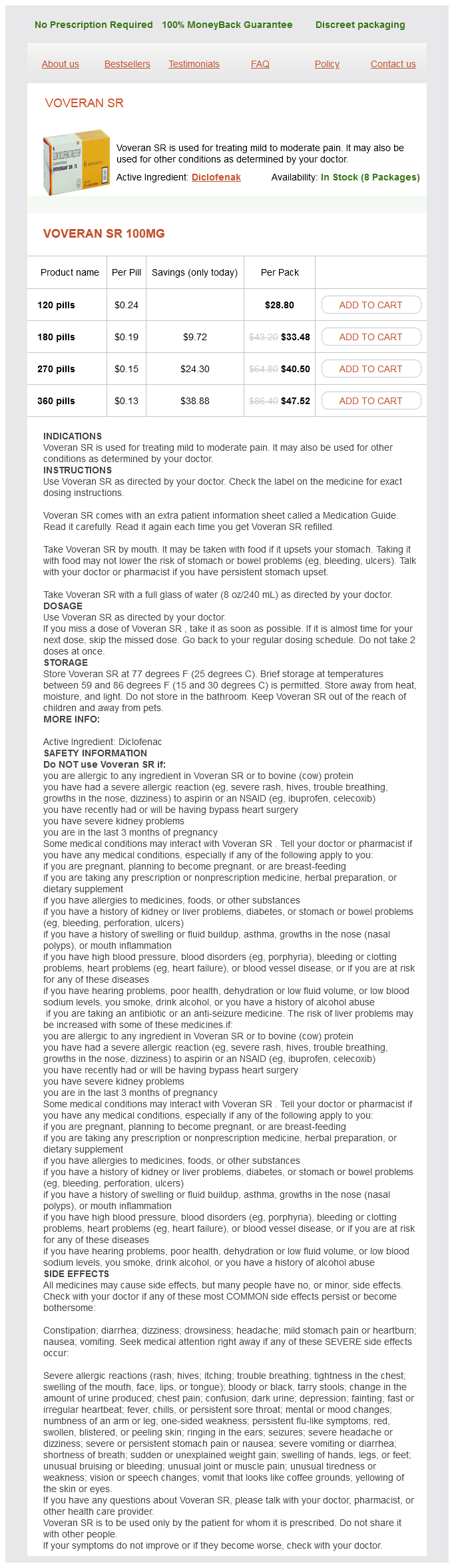
Voveran sr
Voveran sr 100mg
- 120 pills - $28.80
- 180 pills - $33.48
- 270 pills - $40.50
- 360 pills - $47.52
Voveran sr dosages: 100 mg
Voveran sr packs: 120 pills, 180 pills, 270 pills, 360 pills
In stock: 846
Only $0.14 per item
Description
These topical agents are generally safe and useful adjuncts for hemostasis when used for appropriate indications and anatomic sites muscle relaxant otc discount 100 mg voveran sr free shipping. The extent of coagulopathy varies with the type and stage of liver and biliary tract disease. The policy on blood transfusion was changed recently-acceptance of fractionated products of "primary components" was left to the individual believer (Table 44. Improved pathogen detection in donors and pathogen reduction treatments should allow increasingly safer supplies of allogeneic blood products. Proper identifications of the donor unit and patient and careful riskbenefit assessments are essential prior to each component transfusion. Expedited surgical control of bleeding is crucial in improving morbidity and mortality, and there does not appear to be any specific ratio of blood products that can be recommended. The use of thromboelastography/thromboelastometry can be helpful in the selection of hemostatic product(s). Use of factor concentrates in acquired factor deficiency is generally considered to be off-label, but there are some supportive data available for certain indications. The proper indication(s) and site(s) should be considered to reduce untoward complications. Notably, fluid overload or similar cardiac events were more frequent in the plasma group (3% vs. Improved hemostasis was more frequently observed in the 1:1:1 group than in the 1:1:2 group (86. The increased donor exposure was evident in the 1:1:1 group (median 19 U plasma plus platelets vs. This study failed to point to the optimal ratio (or volume) of plasma and platelet transfusion. Fibrinogen or fibrin polymerization was not continuously monitored and cryoprecipitate was seldom used in either group of patients (median, 0 unit). In addition, the incidences of major hemorrhage or cardiac tamponade leading to reoperation were reduced by 50% (P = 0. Thromboelastometry-guided intraoperative haemostatic management reduces bleeding and red cell transfusion after paediatric cardiac surgery. Platelets and plasma were the only 2 products available for hemostatic therapy during the study. Hyperfibrinolysis was evident on thromboelastometry in only 5% of patients, while plasma fibrinolysis markers were elevated in 57% of patients in proportion to the base deficit and hypotension on admission.
Gomphrena paniculata (Suma). Voveran sr.
- Dosing considerations for Suma.
- What is Suma?
- Improving the immune system, cancer and tumors, diabetes, wounds, skin problems, and sexual problems.
- Are there safety concerns?
- How does Suma work?
Source: http://www.rxlist.com/script/main/art.asp?articlekey=96378
The thoracic duct conveys most lymph of the body to the venous system: that from the lower limbs; pelvic cavity; abdominal cavity; left upper limb; and left side of the thorax muscle relaxant flexeril order voveran sr 100 mg with visa, head, and neck-that is, all lymph except that from the right superior quadrant (see the overview of the lymphatic system in the Introduction chapter). It ascends in the posterior mediastinum among the thoracic aorta on its left, the azygos vein on its right, the esophagus anteriorly, and the vertebral bodies posteriorly. The thoracic duct receives branches from the middle and superior intercostal spaces of both sides through several collecting trunks. There are several nodes posterior to the inferior part of the esophagus and more (up to eight) anterior and lateral to it. The posterior mediastinal lymph nodes receive lymph from the esophagus, the posterior aspect of the pericardium and diaphragm, and the middle posterior intercostal spaces. The azygos system exhibits much variation in its origin, course, tributaries, and anastomoses. It ascends in the posterior mediastinum, passing close to the right sides of the bodies of the inferior 8 thoracic vertebrae. In addition to the posterior intercostal veins, the azygos vein communicates with the vertebral venous plexuses that drain the back, vertebrae, and structures in the vertebral canal. The right lymphatic duct is formed by the union of the contralateral partners of the ducts that join the termination of the thoracic duct. The hemi-azygos vein arises on the left side by the junction of the left subcostal and ascending lumbar veins. It ascends on the left side of the vertebral column, posterior to the thoracic aorta as far as the T9 vertebra. Here it crosses to the right, posterior to the aorta, thoracic duct, and esophagus, and joins the azygos vein. The hemi-azygos vein receives the inferior three posterior intercostal veins, the inferior esophageal veins, and several small mediastinal veins. It receives tributaries from veins in the 4th8th intercostal spaces and sometimes from the left bronchial veins. It crosses over the T7 or T8 vertebra, posterior to the thoracic aorta and thoracic duct, where it joins the azygos vein. The left superior intercostal vein, which drains the 1st3rd intercostal spaces, may communicate with the accessory hemi-azygos vein; however, it drains primarily into the left brachiocephalic vein. It is continuous with the superior mediastinum at the sternal angle and is limited inferiorly by the diaphragm. The anterior mediastinum consists of loose connective tissue (sternopericardial ligaments), fat, lymphatic vessels, a few lymph nodes, and branches of the internal thoracic vessels. In unusual cases, this lymphoid organ may extend to the level of the 4th costal cartilages. The apex beat is the impulse that results from the apex of the heart being forced against the anterior thoracic wall when the left ventricle contracts.
Specifications/Details
The anterior and posterior parts of the deltoids are used to swing the limbs during walking spasms when i pee discount 100 mg voveran sr. The deltoid also helps stabilize the glenohumeral joint and hold the head of the humerus in the glenoid cavity during movements of the upper limb. If the deltoid is acting normally, contraction of the middle part of the muscle can be palpated. It can also help extend it from the flexed position, and is an important stabilizer of the humeral head in the glenoid cavity-that is, it steadies the head in its socket. To test the teres major (or the lower subscapular nerve that supplies it), the abducted arm is adducted against resistance. All except the supraspinatus are rotators of the humerus; the supraspinatus, besides being part of the rotator cuff, initiates and assists the deltoid in the first 15° of abduction of the arm. The tendons of the muscles (represented by three fingers and the thumb) blend with the fibrous layer of the capsule of the shoulder joint to form a musculotendinous rotator cuff, which reinforces the capsule on three sides (anteriorly, superiorly, and posteriorly) as it provides active support for the joint. The teres minor is a narrow, elongate muscle that is completely hidden by the deltoid, and is often not clearly delineated from the infraspinatus. The teres minor is most clearly distinguished from the infraspinatus by its nerve supply. The teres minor is supplied by the axillary nerve, whereas the infraspinatus is supplied by the suprascapular nerve (Table 6. Surface Anatomy of Pectoral, Scapular, and Deltoid Regions the clavicle is the boundary demarcating the root of the neck from the thorax. It also indicates the "divide" between the deep cervical and axillary "lymph sheds" (like a mountain range dividing watershed areas): Lymph from structures superior to the clavicles drain via the deep cervical nodes, and lymph from structures inferior to the clavicles, as far inferiorly as the umbilicus, drain via the axillary lymph nodes. This depression overlies the clavipectoral (deltopectoral) triangle-bounded by the clavicle superiorly, the pectoralis major medially, and the deltoid laterally-which may be evident in the fossa in lean individuals. When the arm is abducted and then adducted against resistance, the sternocostal part of the pectoralis major can be seen and palpated. If the anterior axillary fold bounding the axilla is grasped between the fingers and thumb, the inferior border of the sternocostal head of the pectoralis major can be felt. The root of the scapular spine (medial end) is located opposite the tip of the T3 spinous process when the arm is adducted. Grasp the inferior scapular angle with the thumb and fingers and move the scapula up and down. When the arm is abducted, observe that the greater tubercle disappears beneath the acromion and is no longer palpable. The deltoid covering the proximal part of the humerus forms the rounded muscular contour of the shoulder. The teres major is prominent when the abducted arm is adducted and medially rotated against resistance (as when a gymnast stabilizes or fixes the shoulder joint during an iron cross maneuver on the rings).
Syndromes
- Methylmalonic acid blood test
- Endoscopy -- the placement of a camera down the throat to see the extent of burns to the esophagus and the stomach
- There is a skin sore or ulcer over the breast
- Porphyria (several types)
- Confusion
- Are there any muscle spasms?
- Contrast can be given through a vein (IV) in your hand or forearm. If contrast is used, you may also be asked not to eat or drink anything for 4-6 hours before the test.
- Colorectal polyps
Related Products
Usage: q.3h.
Additional information:
9 of 10
Votes: 318 votes
Total customer reviews: 318
Customer Reviews
Milok, 62 years: The deep palmar arch lies approximately 1 cm proximal to the superficial palmar arch. For example, a discarded product is both a quality and operational issue, as is a customer complaint; monitoring these occurrences over a period of time may lead to opportunities for quality improvements and increased operational efficiencies. Bursae are located where tendons rub against bone, ligaments, or other tendons, and where skin moves over a bony prominence. If symptoms persist despite nonweight bearing, then prolongednonweightbearingcanbeattempted.
Delazar, 58 years: The somatic nerves lie laterally (adjacent to the walls), with the vascular structures medial to them. The nerve rests on the ischium and then passes posterior to the obturator internus, quadratus femoris, and adductor magnus muscles. A deep infrapatellar bursa lies between thepatellarligament(just aboveitsinsertion)andthe tibia. Initial studies have been reported in humans with bleeding and who were recently taking an Xa inhibitor.
Rufus, 42 years: Sternocleidomastoid Trapezius Trunks of brachial plexus Fracture of clavicle Coracoclavicular ligament Fractures of Humerus Most injuries of the proximal end of the humerus are fractures of the surgical neck. A characteristic sign is ballooning of the cochlear duct, utricle, and saccule caused by an increase in endolymphatic volume. The ligamentum flavum is disrupted (curved black arrow), and the spinous process is avulsed (straight black arrow). The ankle refers to the narrowest and malleolar parts of the distal leg, proximal to the dorsum and heel of the foot, including the ankle joint.
Redge, 49 years: When viscoelastic testing is normal, surgical bleeding should be considered; however, these tests can also be used to guide transfusion of procoagulant therapies. Acute brachial plexus neuritis (brachial plexus neuropathy) is a neurologic disorder of unknown cause that is characterized by the sudden onset of severe pain, usually around the shoulder. Here the electrode is firmly fixed to the trabeculae carneae in the ventricular wall and placed in contact with the endocardium. The initial pathologic process in the progression of cervical spondylosis is intervertebral disc desiccation.
Hamlar, 37 years: The joints divide the superior appendicular skeleton, and thus the limb itself, into four main segments: shoulder, arm, forearm, and hand. If a low titer product is available this can be considered, as well as volume reducing/ washing to remove the incompatible plasma. Lung Intercostal nerve Collateral branch Intercostal Nerve Block Local anesthesia of an intercostal space is produced by injecting an anesthetic agent around the intercostal nerves between the paravertebral line and the area of required anesthesia. The extracellular portion is a long polypeptide, which has an arginine residue at position 41 and a serine at position 42.
Quadir, 31 years: The role of heparin as an antiinflammatory agent is strengthened when one considers that heparin alone has no effects on coagulation and is found in lower orders of the animal kingdom, such as mollusks, which lack a coagulation system. A thick muscular ridge, the supraventricular crest, separates the ridged muscular wall of the inflow part of the chamber from the smooth wall of the conus arteriosus, or outflow part. The lymphoid tissue is aggregated in certain regions to form masses called tonsils. Amniocentesis: Prior to the use of middle cerebral artery peak systolic velocity, serial amniocentesis was used to determine the severity of hemolysis.
Contact
0673406227
dppsmyanmar@gmail.com