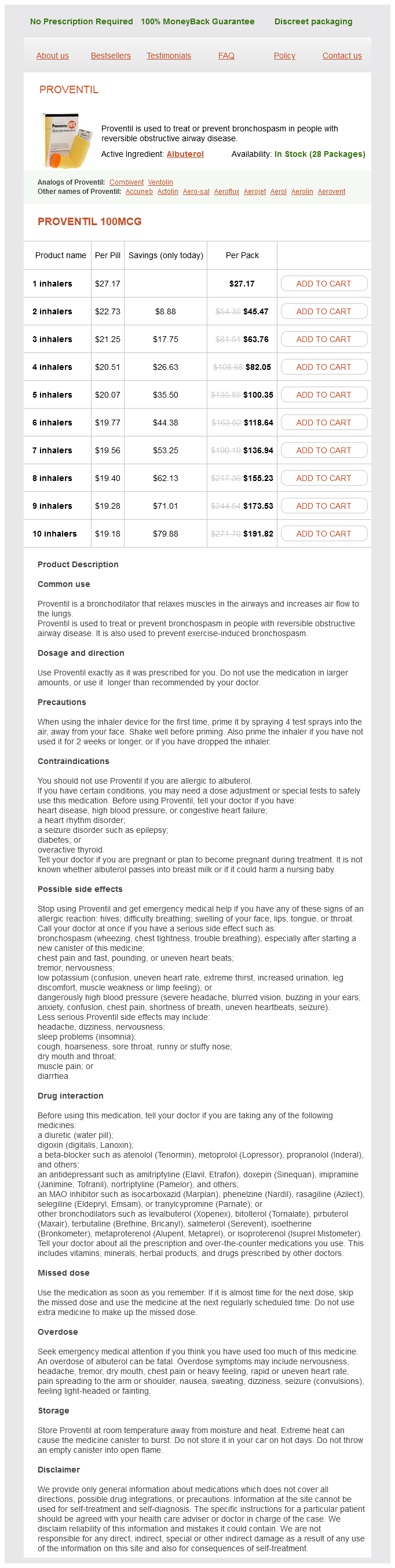
Proventil
Proventil 100mcg
- 1 inhalers - $27.17
- 2 inhalers - $45.47
- 3 inhalers - $63.76
- 4 inhalers - $82.05
- 5 inhalers - $100.35
- 6 inhalers - $118.64
- 7 inhalers - $136.94
- 8 inhalers - $155.23
- 9 inhalers - $173.53
- 10 inhalers - $191.82
Proventil dosages: 100 mcg
Proventil packs: 1 inhalers, 2 inhalers, 3 inhalers, 4 inhalers, 5 inhalers, 6 inhalers, 7 inhalers, 8 inhalers, 9 inhalers, 10 inhalers
In stock: 946
Only $20.38 per item
Description
Retropharyngeal (regardless of laterality); Cervical: unilateral asthma treatment research proventil 100 mcg buy overnight delivery, 6 cm, and above caudal border of cricoid cartilage N2. Superiorly and posteriorly, the mass infiltrates through the longus capitis muscles to involve the entire clivus, anterior occipital bone, and occipital condyles. Intracranially, the tumor infiltrates the bilateral cavernous sinuses with dural thickening and enhancement along the medial walls and floors of middle cranial fossae. Posteriorly, there is retroclival soft-tissue tumor extension with dural enhancement. Anteriorly, the tumor extends to the back of the right nasal cavity, with involvement of the nasal septum, and through the right pterygopalatine fossa to the ipsilateral maxillary sinus. Laterally, the tumor extends into the right parapharyngeal space, with no definite involvement of the right masticator space. Medially, the tumor extends across midline to the left lateral wall of the nasopharynx. Posteriorly, there is involvement of the prevertebral muscles and diffuse involvement of the clivus, bilateral jugular tubercles, and right occipital condyle. There is extension into the right cavernous sinus with cuffing of the petrous portion of the right carotid artery, as well as extension into the right infraorbital fissure and right foramen rotundum. Nasopharyngeal carcinoma Characteristic imaging features: Infiltrative mass centered in the lateral pharyngeal recess. May infiltrate the adjacent soft tissue and skull base, and extend intracranially via direct spread or via skull base foramina. Up to 30 to 45% of patients with perineural spread may be asymptomatic; therefore, imaging assessment for this plays an important role. However, other malignancies such as squamous cell carcinoma, lymphoma, melanoma, basal cell carcinoma, and sarcoma can also show extension along nerves. In practice, most cases of perineural invasion occur in squamous cell carcinoma because it has the highest incidence among all head and neck cancers. Any nerve may serve as a conduit for perineural spread, but the phenomenon is most commonly seen along the maxillary and mandibular divisions of the trigeminal nerve, and the facial nerve. The maxillary nerve innervates many areas in the face, oropharynx, and paranasal sinuses, and may therefore serve as a conduit for many tumors in these areas. Mandibular nerve involvement is often seen in masticator space malignancies and nasopharyngeal carcinoma. It is important to follow the affected nerve in both the antegrade and retrograde directions.
Tantusara (Areca). Proventil.
- Are there safety concerns?
- How does Areca work?
- Dosing considerations for Areca.
- Schizophrenia, glaucoma, or aiding in digestion.
- What is Areca?
- Are there any interactions with medications?
Source: http://www.rxlist.com/script/main/art.asp?articlekey=96956
All the muscles of larynx are supplied by recurrent laryngeal nerve except the cricothyroid muscle asthma definition key 100 mcg proventil buy overnight delivery. Sensory supply to laryngeal mucosa above vocal cords is by internal laryngeal nerve and below the vocal cords by recurrent laryngeal nerve. All intrinsic muscles of larynx are supplied by the recurrent laryngeal nerve except cricothyroid which is supplied by the external laryngeal nerve. This is a question about posterior cricoarytenoid muscle, which is the only abductor of vocal cords in larynx. The diagram shows a transverse view of larynx, with muscles attached to various cartilages and named accordingly. C · · · · · · Trachea Trachea is a part of respiratory tube, beginning below the larynx (level with the sixth cervical vertebra), and ends at the carina (at the level of the disc between T4-5 vertebra, opposite the sternal angle). The extent of trachea varies as follows: C6 to T4 in cadaver placed in supine position. Note: Trachea lies in the midline but point of bifurcation is usually to the right side. Structure: A long tube formed of cartilage and fibromuscular wall, and lined internally by respiratory mucosa. The anterolateral portion of the trachea consists of 1620 U-shaped superimposed incomplete rings of hyaline cartilage and posteriorly lies the trachealis muscle. The last tracheal ring merges into the incomplete rings at the origin of each principal bronchus; the bifurcation is marked by a cartilaginous spur, the carina, which can be observed by bronchoscopy as a raised ridge of tissue in the sagittal plane. Arteries: Branches of the inferior thyroid arteries and their anastomosis with bronchial arteries. It commences at the lower border of the larynx, level with the sixth cervical vertebra. Inside the trachea at the level of the fifth thoracic vertebra (T5) there is a cartilaginous ridge known as the carina of trachea which runs across from the front to the back of the trachea and marks the point of bifurcation into the right and left primary bronchi. The carina is opposite the sternal angle and can be positioned up to two vertebrae lower or higher, depending on breathing. The answer depends upon the statements mentioned in the standard textbooks and it is advisable not to use personal logic and imagination. Lower border of T4 · · · · · · · the most appropriate option has been taken as the answer. Trachea bifurcates into bronchi at the level of the disc between T4-5 vertebra, opposite the sternal angle. Carina (at tracheal bifurcation) is located about 25 cm from the incisor teeth and 30 cm from the nostrils.
Specifications/Details
The medial area is covered by fibroadipose tissue that usually contains the sciatic bursa of gluteus maximus asthma definition 6000 generic proventil 100 mcg buy, which supports the body in sitting. The upper and lower borders of gluteus maximus muscle are indicated by thick red lines Sacrotuberous ligament (runs from the sacrum to the ischial tuberosity) and sacrospinous ligaments (runs from the sacrum to the ischial spine) convert the greater and lesser sciatic notches of the hip bone into greater and lesser sciatic foramina, the two important exits from the pelvis. Sacrotuberous ligament is a broad band of fibrous tissue which extends from sides of the sacrum and coccyx to the medial side of the ischial tuberosity. The lowest fibres of gluteus maximus are attached to it and the lower part of the ligament continue into the tendon of biceps femoris. The coccygeal branches of the inferior gluteal artery, the perforating cutaneous nerve and filaments of the coccygeal plexus pierce the ligament. Sacrospinous ligament extends from the ischial spine to the lateral margins of the sacrum and coccyx anterior to the sacrotuberous ligament. Greater sciatic foramen is bounded anterosuperiorly by the greater sciatic notch, posteriorly by the sacrotuberous ligament and inferiorly by the sacrospinous ligament and ischial spine. Piriformis muscle pass through it, above which the superior gluteal vessels and nerve leave the pelvis. Other important structures as they exit the pelvic cavity to enter the gluteal and thigh regions: Superior gluteal vein, artery, and nerve; inferior gluteal vein, artery, and nerve; sciatic nerve. Adductor magnus · Posterior (hamstring) part of adductor magnus takes origin from the ischial tuberosity. Origin of semimembranosus from superolateral area · Semi-membranosus arises by a long, flat tendon from a superolateral impression on the ischial tuberosity. Obturator internus muscle · It is the tendon (not muscle) of obturator internus, which passes through lesser sciatic notch. Nerve to obturator internus · Pyriformis muscle pass through greater sciatic notch. Coccygeal nerve · the sacrotuberous ligament is pierced by the coccygeal branches of the inferior gluteal artery, the perforating cutaneous nerve and filaments of the coccygeal plexus. It shows the following features: Head forms about two-thirds of a sphere and is directed medially, upward, and slightly forward to fit into the acetabulum. It has a depression in its articular surface, the fovea capitis femoris, to which the ligamentum capitis femoris is attached. It is separated from the shaft in front by the intertrochanteric line, to which the iliofemoral ligament is attached. It provides an insertion for the gluteus medius and minimus, piriformis, and obturator internus muscles.
Syndromes
- Farsightedness
- Tetrabenazine, a medicine to treat excessive movement disorder
- The joint appears warm and red. It is usually very tender and swollen (it hurts to put a sheet or blanket over it).
- Is there any history of joint dislocation, difficulty walking, or difficulty using the arms?
- Drink plenty of fluids every day, even when you are well. Drink more when the weather is hot or you are exercising.
- Commonly has imaginary playmates
- Breathing support (possibly artificial respiration)
- How much of the drug was taken?
Related Products
Usage: q.i.d.
Additional information:
8 of 10
Votes: 312 votes
Total customer reviews: 312
Customer Reviews
Josh, 31 years: Each maxillary molar generally has three roots, and each mandibular molar has two roots. Floor is contributed by the palatine process of the maxilla and the horizontal plate of the palatine bone. The enhancing mass (arrow; a) has a small contiguous enhancing focus that extends into the hypotympanum. Predisposing factors include druginduced liver toxicity from inhalational anesthetics, intraoperative or perioperative hypotension with ischemic liver injury, blood transfusions, total parenteral nutrition, antimicrobials/antifungals, and sepsis.
Hurit, 61 years: Arterial Supply Thoracic wall receive their blood supply from the internal thoracic artery (either directly or via the musculophrenic artery), the superior intercostal artery (from the costocervical trunk), superior thoracic artery (from the axillary artery), descending thoracic aorta, and the subcostal artery. Stackable carbon fiber cages for thoracolumbar interbody fusion after corpectomy: long-term outcome analysis. Long-term, progression-free survival rate is greater than 90% with localized pure germinomas after radiation therapy. In this procedure, the periorbita is dissected free from the medial orbital wall and the orbital floor.
Contact
0673406227
dppsmyanmar@gmail.com