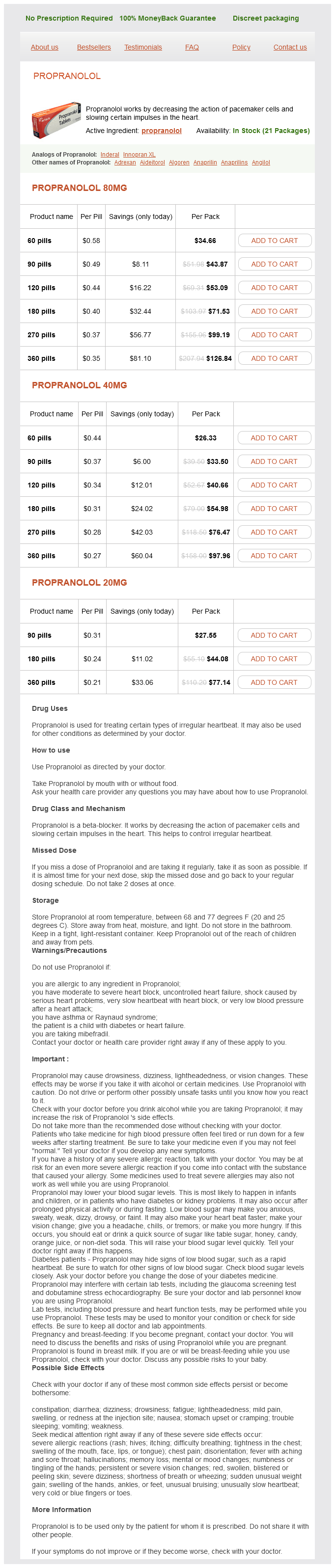
Propranolol
Propranolol 80mg
- 60 pills - $34.66
- 90 pills - $43.87
- 120 pills - $53.09
- 180 pills - $71.53
- 270 pills - $99.19
- 360 pills - $126.84
Propranolol 40mg
- 60 pills - $26.33
- 90 pills - $33.50
- 120 pills - $40.66
- 180 pills - $54.98
- 270 pills - $76.47
- 360 pills - $97.96
Propranolol 20mg
- 90 pills - $27.55
- 180 pills - $44.08
- 360 pills - $77.14
Propranolol dosages: 80 mg, 40 mg, 20 mg
Propranolol packs: 60 pills, 90 pills, 120 pills, 180 pills, 270 pills, 360 pills
In stock: 993
Only $0.23 per item
Description
Allantosis-urinary bladder 2 arteries 100 blocked cheap 20 mg propranolol, vagina, urethra, secretory cells of the prostate and urethral glands. Coelomic wall epithelium Walls of intra-embryonic coelom Primitive pericardium - myocardium, parietal pericardium pericardioperitoneal canals - visceral, parietal and mediastinal pleura, pleuroperitoneal membranes contributing to diaphragm Splanchnopleuric epithelium - visceral peritoneum of stomach, peritoneum of lesser and greater omental, faicliform ligament, lienorenal and gastrosplenica ligaments Somatopleuric epithelium - parietal peritoneum Primitive peritoneal cavity Splanchnopleuric epithelium - visceral peritoneal covering of mid- and hindgut, the mesentery, transverse and sigmoid mesocolon. Pronephros, epithelial lining of mesonephric ducts, vas deferens, epididymis, seminal vesicles, ejaculatory duct, ureters, vesical trigone Mullerian ducts, epithetical lining of uterine tubes, body and cervix of uterus, vagina, broad ligament of uterus. Germinal epithelium of gonad (note the germ cells are not included on this chart because of their early sequestration into the extraembryonic tissues) Germinal epithelium forming cortex of suprarenal gland. Paraxial mesenchyme (somites and somitomeres) Sclerotome - vertebrae and portions of the neurocranium, axial skeleton Myotome - all voluntary muscles of the head, trunk and limbs. Intermediate mesenchyme - connective tissue of gonads, mesonephric and metanephric nephrons, samooth muscle and connective tissues of the reproductive tracts Septum transversum - epicardium, fibrous pericardium, portion of diaphragm, oesophageal mesentery, sinusoids of liver, tissue within lesser omentum and falciform ligament. Lateral plate mesenchyme Splanchnopleuric layer - smooth muscle and connective tissues of respiratory tract and associated glands. Smooth muscle and connective tissues of intestinal tract, associated glands and abdominal mesenteries. Smooth muscle and connective tissue of blood vessels (also see below) Somatopleuric layer - appendicular skeleton, connective tissue of limbs and trunk, including cartilage, ligaments and tendons. Angiogenic mesenchyme Endocardium of heart, endothelium of blood and lymphatic vessels, vessels of choroid plexus, sinusoids of liver and spleen, circulating blood cells, microglia, tissue macrophages. Olfactory receptor cells and olfactory epithelium Epithelial walls of the membranous labyrinth, the cochlear organ of corti. Lens of the eye Enamel organs of the teeth Cranial structures Secretory and duct-lining cells of the lacrimal, nasal, labial, palaver, oral and salivary glands. Epithelial lining of the external acoustic mealsus and external epithelium of the lymphatic membrane. Epithelial lining of the lacrimal canaliculi and nasolacrimal duct Epithelial lining of the paranasal sinuses, lips, cheeks, gums and palate. Ependyma lining the cerebral verticles, aqueduct and central canal of brain and spinal cord, tarycytes, cells covering the choroid plexuses, circumventricular cells. Sensory neurones of the spinal dorsal root ganglia and their peripheral sensory receptors Satellte cells in all sensory ganglia Sympathetic ganglia and plexuses: neurones and satellte cells Parasympathetic ganglia and plexuses: neurones and satellte cells Enteric plexuses: neurones and gllal cells Schwann cells of the all the peripheral nerves Medulla of the suprarenal glands. Calcilonin-producing cells (C cells) Melanocytes Mesenchymal derivatives in the head Frontal, parietal, squamous temporal, nasal, vomer, palatine bones, maxillae and mandile Meninges 55 Self Assessment and Review of Anatomy Surface ectoderm epithelium Epidermal structures Most of the cutaneous epidermal cells, the secretary, duct-lining and myoepithelial cells of the sweat, sebaceous and mammary glands Hair and nails. Proctodeal epithelium and epithelium of the terminal malo urethra Neural plate epithelium Neural crest Choroid and sclera of eye Connective tissue of lacrimal, nasal, labial, pataline, oral and salivary glands Dentine of teeth Connective tissues of thyroid glands and of the pharyngeal pouches, i. Somites give rise to the vertebrae of the vertebral column, rib cage, and part of the occipital bone; skeletal muscle, cartilage, tendons, and skin dermis (of the back). Somites are differentiated into: · Dermomyotome: Form skeletal muscles and dermis. Each lateral plate splits horizontally into the dorsal somatic (parietal) mesoderm, which underlies the ectoderm, and the ventral splanchnic (visceral) mesoderm, which overlies the endoderm.
Aristolochia serpentaria (Aristolochia). Propranolol.
- Dosing considerations for Aristolochia.
- Sexual arousal, convulsions, immune stimulation, promoting menstruation, colic, gallbladder cramps, arthritis, gout, rheumatism, eczema, weight loss, and wound treatment.
- Are there safety concerns?
- What is Aristolochia?
- How does Aristolochia work?
Source: http://www.rxlist.com/script/main/art.asp?articlekey=96579
Spinal accessory nerve roots arise from spinal cord C1-5 cardiovascular means purchase propranolol 80 mg overnight delivery, form a trunk that ascends in the vertebral canal, enter foramen magnum and join the cranial part. Both pass through the jugular foramen, cranial accessory fibres join the vagus nerve (vagus accessory complex) whereas, the spinal accessory nerve supplies sternocleidomastoid muscle and trapezius. Cranial accessory nerve (neuron bodies in nucleus ambigus) fibres are carried by vagal branches to supply muscles of palate, pharynx and larynx. Stylopharyngeus muscle is supplied by the glossopharyngeal nerve, fibres arising from nucleus ambigus. Receives contributions from vagus nerve carrying cranial accessory nerve component b. Stylopharyngeus · · · · · · Stylopharyngeus is innervated by the glossopharyngeal nerve and not the accessory nerve. All the other muscles are supplied by the vago-accessory complex / pharyngeal plexus. The pharyngeal branch of the vagus supplies all the muscles of the pharynx (excluding stylopharyngeus, which is supplied by the glossopharyngeal nerve) and of the soft palate (excluding tensor veli palatini, which is supplied by the mandibular division of the trigeminal via the nerve to medial pterygoid). It emerges from the upper part of the inferior vagal ganglion and consists mainly of filaments derived from the cranial accessory nerve: almost all the neuronal cell bodies are in the nucleus ambiguus. Stylopharyngeus muscle is the only muscle developing in the third pharyngeal arch and the only muscle to be supplied by the glossopharyngeal nerve. Most of the muscles of the palate and pharynx develop in the fourth pharyngeal arch and are supplied by the cranial part of accessory nerve, whose axons are carried by the vagus nerve through the pharyngeal plexus to the muscles. Tensor tympani muscle develops in first pharyngeal arch and is supplied by the mandibular nerve (trigeminal). Vagus nerve carry axons of cranial accessory nerve (vagus accessory complex) and gives pharyngeal branches to pharyngeal plexus and the plexus itself sends these axons to muscles of palate and pharynx. Cranial part of accessory nerve supplies most of the pharyngeal muscles with few exceptions. Stylopharyngeus develops in third pharyngeal arch and supplied by glossopharyngeal nerve. Most of the muscle of palate are supplied by cranial accessory nerve (via vagus accessory complex) except few exceptions. Tensor veli palate develops in first pharyngeal arch and supplied by mandibular branch of trigeminal nerve. Tensor veli palati 382 Head and Neck Hypoglossal nerve Hypoglossal Nerve is given by the medulla oblongata ventrally in the preolivary sulcus and passes through the hypoglossal It loops around the occipital artery and the carotid bifurcation (in carotid triangle) to pass between the carotids and It runs deep to the digastric posterior belly and stylohyoid muscles to enter the submandibular triangle. It enters the mouth by passing above the greater horn of the hyoid bone between the middle pharyngeal constrictor and After crossing the loop of the lingual artery a little above the tip of the greater cornu of the hyoid, it inclines upwards and Between mylohyoid and hyoglossus, the hypoglossal nerve lies below the deep part of the submandibular gland, the It then passes on to the lateral aspect of genioglossus, continuing forwards in its substance as far as the tip of the tongue. It supplies motor fibers to all of the intrinsic and extrinsic muscles of the tongue except the palatoglossus (which is supplied It carries sensory fibers from C1 to supply the cranial dura mater through the meningeal branch to supply the upper root of the ansa cervicalis and the nerve to both the thyrohyoid and geniohyoid muscles.
Specifications/Details
The latter also commonly arises due to pain coronary heart xmas cheap propranolol 40 mg buy on line, especially in the setting of higher stage locally advanced disease. Knowledge of the clinical location of the tumor relative to the anal verge is invaluable in obtaining diagnostic images. Defining the peritoneal reflection has been shown to be accurate for surgical planning. The continued pursuit of a means to identify the dentate line, instead of relying on the anal verge, may result in a better determination of the appropriateness for a sphincter-saving procedure. Lastly, the ability to define the complete responder with negative nodal status will reduce the number of patients undergoing major surgery that results in detrimental long-term change in quality-of-life measures for no benefit in the ultimate cure of the cancer. International preoperative rectal cancer management: staging, neoadjuvant treatment, and impact of multidisciplinary teams. High rate of positive circumferential margins following rectal cancer surgery: a call to action. Watch and wait approach following extended neoadjuvant chemoradiation for distal rectal cancer: are we getting closer to anal cancer management Effectiveness of preoperative staging in rectal cancer: digital rectal examination, endoluminal ultrasound or magnetic resonance imaging Magnetic resonance tumor regression grade and residual mucosal abnormality as predictors for pathologic complete response in rectal cancer postneoadjuvant chemoradiotherapy. Rectal cancer with complete clinical response after neoadjuvant chemotherapy, surgery, or "watch and wait. A single-centre experience of chemoradiotherapy for rectal cancer: is there potential for nonoperative management Wait-and-see policy for clinical complete responders after chemoradiation for rectal cancer. Nonoperative management of rectal cancer with complete clinical response after neoadjuvant therapy. The procedures can be done in the office setting with portable equipment and no sedation. Minimal preparation is required, tolerance by patients is high, and the results are readily available. Patients are instructed to use one or two enemas prior to the office visit because fecal material within the rectum causes image artifacts. Examinations are performed in the kneeling prone or left lateral position, depending on practitioner preference. Any residual fecal material and enema effluent can be suctioned out to decrease image artifacts. The distance from the tumor to the anal verge is measured, the location of the tumor on the rectal wall and the size of the rectal lumen through the tumor are noted.
Syndromes
- Percutaneous means through the skin. Most kidney biopsies are done this way.
- To help control high blood pressure in someone who has problems with the blood supply to their kidney
- Fever
- Eat slowly and chew food thoroughly.
- Tingling or numbness of the skin
- Advocacy groups -- especially to help you find the best provider for a specific chronic condition or disability
- Sinusitis - acute
- · Avoid raw or undercooked meat and fish.
- You are given medicine to help you relax.
- Deformities in the spine
Related Products
Usage: p.r.n.
Additional information:
8 of 10
Votes: 264 votes
Total customer reviews: 264
Customer Reviews
Asam, 51 years: In the presence of a hepatic flexure or proximal colon tumor, the omentum may be involved and should remain attached to the colon to ensure an en bloc resection. Safety of performing a delayed anastomosis during damage control laparotomy in patients with destructive colon injuries. The splenic vein is dealt with in the same manner, and the splenectomy is completed.
Snorre, 23 years: The 2-year overall survival for patients with an hdb less than 25% versus greater than 25% was 42% and 0%, respectively. Before passing the ileum through the abdominal wall, proper orientation is ensured and the distal end is marked with a suture to prevent maturation of the incorrect segment after the abdominal incision has been closed. Haematogenous metastatic patterns in colonic carcinoma: an analysis of 1541 necropsies.
Contact
0673406227
dppsmyanmar@gmail.com