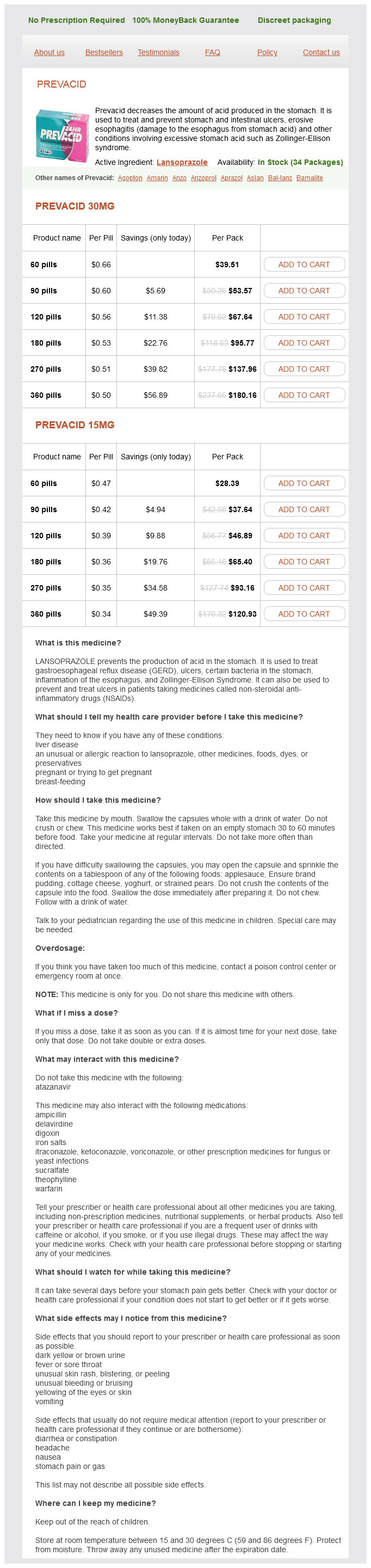
Prevacid
Prevacid 30mg
- 60 pills - $39.51
- 90 pills - $53.57
- 120 pills - $67.64
- 180 pills - $95.77
- 270 pills - $137.96
- 360 pills - $180.16
Prevacid 15mg
- 60 pills - $28.39
- 90 pills - $37.64
- 120 pills - $46.89
- 180 pills - $65.40
- 270 pills - $93.16
- 360 pills - $120.93
Prevacid dosages: 30 mg, 15 mg
Prevacid packs: 60 pills, 90 pills, 120 pills, 180 pills, 270 pills, 360 pills
In stock: 510
Only $0.36 per item
Description
These studies led to the emergence of a model system for cell adhesion and signal transduction [10] gastritis ct cheap prevacid 15 mg buy on-line. This was also the beginning of the understanding of the Wnt/catenin pathway and its role in complex cellular processes such as cellcell adhesion, mitogenesis, motogenesis and morphogenesis in the vertebrates. The next several years focused on the discovery of various components of this pathway that improved our understanding of the regulation of this complex signaling cascade in normal physiology and disease. Many new components and interactions as well as expanding list of target genes are being identified. Also, research is focused on their role in regulation of this pathway in health and disease. Presently, the role of Wnt/catenin pathway is well established in vertebrates in development, tissue homeostasis, and carcinogenesis [11, 12]. Availability of conditional knockouts to overcome embryonic lethality has been key to understanding a more ubiquitous role of catenin and other Wnt components in the development of many organs such as kidneys, lungs, brain, limbs, muscles, and skin [1419]. The role of Wnt/catenin signaling in liver pathobiology has gained significant importance in the last 1520 years. This article will highlight the role of the Wnt/catenin signaling pathway and of catenin independent of the canonical Wnt pathway in liver physiology and pathobiology (Table 46. Although the most characterized signaling downstream of the the Liver: Biology and Pathobiology, Sixth Edition. In this, a broad overview of the various signaling cascades activated downstream of Wnt are discussed. However, Wnt proteins, in order to be biologically active, need to undergo specific posttranslational modifications. Several target genes of the pathway have now been identified, which are stage and tissuespecific, and the liver related targets will be discussed throughout the text. Understandably, not all Wnt signaling converges on catenin and various WntFzd or WntnonFzd interactions can lead to activation of alternate signaling pathways that are briefly discussed here. It should be noted that some Wnts that were classically dubbed to be noncanonical have now been shown to activate or inactivate catenin based on interaction with specific receptors and the final outcome of a particular signaling is contextdependent. An example is that of Wnt5a, which can activate or inhibit catenin based on the receptor it binds [32]. On the right, Wnt is glycosylated and palmitoylated by Porcupine (Porcn) in the endoplasmic reticulum and is bound to Wntless (Wls) in the Golgi apparatus to be transported to the membrane for secretion. If it binds to Fzd4, it can activate catenin while if it binds to Ror2 or Fzd2, it can inactivate catenin through Wnt/calcium pathway or other mechanisms.
Blue Green Algae (Blue-Green Algae). Prevacid.
- What is Blue-green Algae?
- Attention deficit-hyperactivity disorder (ADHD), premenstrual syndrome (PMS), diabetes, to stimulate the immune system, fatigue, anxiety, depression, memory, energy, high cholesterol, heart disease, precancerous mouth lesions (oral leukoplakia), wound healing, weight loss, digestion, tics or twitching of the eyelids (called blepharospasm or Meige syndrome), and as a source of dietary protein, vitamin B12, and iron.
- How does Blue-green Algae work?
- Are there any interactions with medications?
- Dosing considerations for Blue-green Algae.
- Weight loss.
- Treating precancerous mouth lesions.
- Are there safety concerns?
Source: http://www.rxlist.com/script/main/art.asp?articlekey=96887
Work from our laboratory further elucidated the requirement for intact Yap activity for liver development: zebrafish embryos mutant for yap exhibited small hypoplastic livers with decreased proliferative index gastritis symptoms upper back pain order prevacid 15 mg line. The central role of Yapdependent nucleotide production for normal liver development is emphasized by the potential for external nucleotides to partially rescue liver development [23]. Interestingly, concomitant deletion of Yap and Taz in the liver leads to an increased organ size at two months of age, with evidence of increased cell proliferation [28]. What has yet to be determined is what metabolic changes, ranging from glucose and glutamine concentrations to reactive oxygen species concentration and hypoxia, are experienced in the developing embryo, leading to modulation of these pathways and their downstream impact on hepatobiliary development. Given the central role of the liver in metabolic homeostasis, liver injury leads to significant alterations in glucose, nitrogen, and lipid and bile salt metabolism [3133]. In particular, mice undergoing partial hepatectomy develop significant hypoglycemia due to the loss of liver tissue normally involved in gluconeogenesis [34] as well as steatosis [33, 35, 36]. Further, carbohydrate restriction prior to liver injury significantly impairs liver repair [37], however, alleviating the hypoglycemic response after liver injury with dextrose infusion delayed liver regeneration [38]. The bestdescribed metabolites affecting liver regeneration may be bile acids: intrahepatic levels of bile acids increase rapidly after resection [43, 44]. Similarly, deletion of the bile acid membrane transporter Mrp3 (Abcc3) in mice impaired liver regeneration after partial hepatectomy due to lower portal blood concentrations of bile acids [48]. These studies demonstrate that changes in metabolism occur after liver injury and can directly affect liver regeneration. With the exception of bile salts, however, it is still largely unclear how the liver senses and responds to metabolic alterations occurring after liver injury, but the involvement of central nutrient sensing and regulating pathways strongly suggest that the process of liver regeneration is at least in part regulated by metabolic changes occurring as a consequence of liver injury. Interestingly, these changes appear to be independent of metabolic changes in glucose and lipid metabolism well described after partial hepatectomy. Further, Lkb1 regulates the spindle integrity during mitosis, thereby affecting the ploidy state and contributing to genomic integrity of the regenerating liver. Nevertheless, its role in liver regeneration is less well defined: after partial hepatectomy, treatment with rapamycin decreases Brdu incorporation and cell proliferation in mice and rats [21, 56]. Treatment with rapamycin, its analogs, or genetic deletion of S6K results in a delay in S phase entry and decreased hepatocyte proliferation [21, 58]. Liver kinase B1 regulates hepatocellular tight junction distribution and function in vivo. Ribosomal protein S6 phosphorylation and function during late gestation liver development in the rat. Yap regulates glucose utilization and sustains nucleotide synthesis to enable organ growth. Yap reprograms glutamine metabolism to increase nucleotide biosynthesis and enable liver growth. The Hippo signaling functions through the Notch signaling to regulate intrahepatic bile duct development in mammals.
Specifications/Details
This hypocontractility occurs largely due to the presence of excessive vasodilator molecules gastritis medicine cvs purchase prevacid 15 mg free shipping. In arteries of the splanchnic circulation, it has been shown that vasodilators increase and vasoconstrictors decrease. This syndrome is characterized by increased cardiac index, decreased systemic vascular resistance, and decreased mean arterial pressure and eventually leads to multiple organ failure frequently observed in chronic liver disease. Neural factors Neural factors are also thought to be involved in the dysfunction of vascular tone in portal hypertension, especially through the sympathetic nervous system [101, 107, 108]. It is reported that sympathetic nerve atrophy/regression observed in mesenteric arterial beds of portal hypertensive rats leads to vasodilation and/or hypocontractility of those arterial beds [109, 110]. The role of neural factors in decreased contractile responses remains to be fully elucidated. The systemic circulation and the heart Although arterial vasodilation in the splanchnic circulation is an essential initiating factor, hyperdynamic circulation does not occur without expansion of plasma volume and the development of portosystemic collaterals [125, 126]. In cirrhotic patients, blood and plasma volumes are elevated, but are not evenly distributed among vascular areas [127]. For example, arterial blood volumes in the heart, lungs, and central arterial tree are decreased compared to those in the splanchnic circulation, resulting in central hypovolemia. Decreased blood volumes together with arterial hypotension lead to baroreceptor activation of potent vasoconstriction systems such as the sympathetic nervous system and the renin angiotensin aldosterone system [128], resulting in water retention and plasma volume expansion. The combination of an expanded plasma volume and a reduction in peripheral vascular resistance leads to an increase in cardiac output. When portal hypertension persists, the heart results in a high cardiac output syndrome: initial compensation occurs according to the degree of individual cardiac output, followed by some degree of cardiac insufficiency. The cardiac index is usually higher than normal (greater than 4 L min-1 m2) but insufficient to maintain arterial pressure in the face of progressive arterial vasodilation [5]. Importantly, high cardiac output failure is reversible once the initial cause leading to the high cardiac output is treated. This reversal has also been observed in patients with cirrhosis after liver transplantation [129, 130]. Structural changes of arteries the thinning of arterial walls is observed in the splanchnic and systemic circulations of rats with cirrhotic livers [111, 112]. While this arterial wall thinning can be a consequence of the hyperdynamic circulation, it may also sustain arterial vasodilation and exacerbate portal hypertension [5, 6]. Collateral vessel formation Portosystemic collateral vessels develop in response to an increase in portal pressure. These collateral vessels develop in an attempt to decompress the hypertensive portal system and are formed through the opening of preexisting vessels or angiogenesis [113, 114]. However, these collateral vessels are also known to cause serious complications, including variceal bleeding and hepatic encephalopathy [5, 115, 116]. A change in portal pressure is thought to be detected first by the intestinal microcirculatory bed, followed by arteries of the splanchnic circulation [51].
Syndromes
- Difficulty sleeping due to the itching that occurs during the night
- Impotence
- This test can be used to diagnose the infection and confirm that it has been cured after treatment.
- Dark stools
- White blood cells (lymphocytes) on a thyroid biopsy
- Get medical attention.
- Dizziness or vertigo
Related Products
Usage: ut dict.
Additional information:
9 of 10
Votes: 99 votes
Total customer reviews: 99
Customer Reviews
Hurit, 32 years: There are at least two other subdominant epitopes, with the second usually designated as either "d" or "y" and the third being either "w" or "r. Tolllike receptor 2 and palmitic acid cooperatively contribute to the development of nonalcoholic steatohepatitis through inflammasome activation in mice. Race the role of race and ethnicity in the outcome of chronic hepatitis C is controversial.
Hjalte, 50 years: In contrast, amino acid substitutions were not detected in patients in whom infection resolved spontaneously, including one of two patients infected from a single source who developed divergent outcomes of infection [254]. Tachyzoites were present in tissue sections of parasites developing in muscle, heart, liver, spleen, kidney, pancreas, reproductive organs, eyes, and brain (Sanders et al. The parasite appears to possess transporters capable of transporting ornithine, putrescine, and other polyamines (Chaudhary, 2005; Chaudhary et al.
Vatras, 56 years: An alternative approach utilizes E1Bmutant adenoviruses that are capable of replicating in cells that lack active p53, which includes many tumor cells [105]. In one such study, cases of transfusionrelated nonA, nonB hepatitis identified from five prospective transfusionrelated hepatitis studies were combined and matched 2:1 with persons transfused without hepatitis [237]. Broadly neutralizing antibodies protect against hepatitis C virus quasispecies challenge.
Contact
0673406227
dppsmyanmar@gmail.com