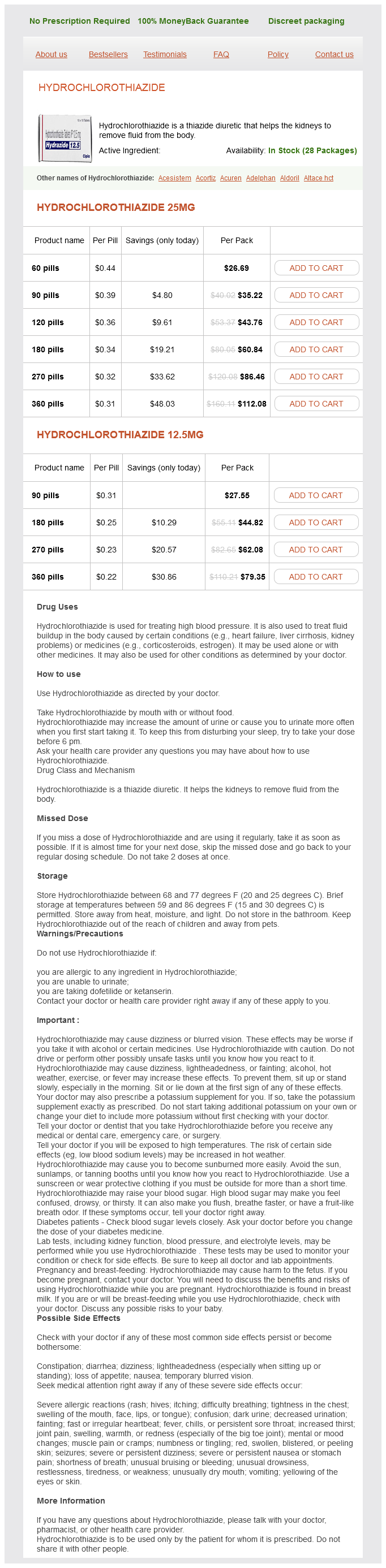
Hydrochlorothiazide
Hydrochlorothiazide 25mg
- 60 pills - $26.69
- 90 pills - $35.22
- 120 pills - $43.76
- 180 pills - $60.84
- 270 pills - $86.46
- 360 pills - $112.08
Hydrochlorothiazide 12.5mg
- 90 pills - $27.55
- 180 pills - $44.82
- 270 pills - $62.08
- 360 pills - $79.35
Hydrochlorothiazide dosages: 25 mg, 12.5 mg
Hydrochlorothiazide packs: 60 pills, 90 pills, 120 pills, 180 pills, 270 pills, 360 pills
In stock: 996
Only $0.23 per item
Description
The long axis of the lobule is traversed by the terminal hepatic venule (central vein) blood pressure joint pain 12.5 mg hydrochlorothiazide order overnight delivery, which receives blood from the hepatic sinusoids. Note that a wedge of the tissue has been removed from the lobule for better visualization of the terminal hepatic venule. Interconnecting sheets or plates of hepatocytes are disposed in a radial pattern from the terminal hepatic venule to the periphery of the lobule. The liver acinus is lozenge-shaped and represents the smallest functional unit of the hepatic parenchyma. The short axis of the acinus is defined by the terminal branches of the portal triad that lie along the border between two classic lobules. The long axis of the acinus is a line drawn between the two central veins closest to the short axis. This photomicrograph shows a cross-section of a pig liver lobule stained by the Mallory-Azan method to visualize connective tissue components. Note the relatively thick interlobular connective tissue (stained blue) surrounding the lobules. The terminal hepatic venule (central vein) is visible in the center of the lobule. Note that in contrast to the pig liver, the lobules of the human liver lack connective tissue septa. The boundaries of a lobule can be approximated, however, by drawing a line (dashed line) from one portal canal to the next, thus circumscribing the lobule. The outlines of a classic hepatic lobule, portal lobule, and liver acinus are visible on this section of the liver tissue. Note that the hexagonal-shaped classic lobule (red) has the terminal hepatic venule (central vein) at the center of the lobule and the portal canals containing portal triads at the peripheral angles of the lobule. The triangular portal lobule (green) has a portal canal at the center of the lobule and terminal hepatic venules (central veins) at the peripheral angles of the lobule. A diamondshaped liver acinus (multicolor) has distributing vessels at the equator and terminal hepatic venules (central veins) at each pole. It consists of adjacent sectors of neighboring hexagonal fields of classic lobules partially separated by distributing blood vessels. The zones, marked 1, 2, and 3, are supplied with blood that is richest and most nutrient-oxygenated in zone 1 and least so in zone 3. The terminal hepatic venules (central veins) in this interpretation are at the pointed edges of the acinus instead of in the center, as in the classic lobule. The portal triads (terminal branches of the portal vein and hepatic artery) and the smallest bile ducts are shown at the corners of the hexagon that outlines the cross-sectioned profile of the classic lobule. This concept allows a description of the exocrine secretory function of the liver comparable to that of the portal lobule.
Kana (Indian Long Pepper). Hydrochlorothiazide.
- Are there safety concerns?
- What is Indian Long Pepper?
- Headache, toothache, asthma, bronchitis, cholera, coma, cough, diarrhea, epilepsy, fever, stomachache, stroke, indigestion, menstrual disorders, and other conditions.
- Dosing considerations for Indian Long Pepper.
- How does Indian Long Pepper work?
- Are there any interactions with medications?
Source: http://www.rxlist.com/script/main/art.asp?articlekey=96385
The liver is the major blood-forming organ in the fetus during the second trimester prehypertension treatment 12.5 mg hydrochlorothiazide purchase with visa. The third or bone marrow phase of fetal hemopoiesis and leukopoiesis involves the bone marrow (and other lymphatic tissues) and begins during the second trimester of pregnancy. Cytokines (including hemopoietic growth factors) may and do act individually and severally at any point in the process from the first stem cell to the mature blood or connective tissue cell. If committed to enter the mast cell lineage, the basophil/mast cell progenitor cell migrates to the spleen where it differentiates into a mast cell progenitor cell. After further differentiation in the spleen, it migrates to the intestine to become a mast cell precursor. Although it is difficult to discern, these cells are located between developing liver cells and the wall of the vascular sinus. Monophyletic Theory of Hemopoiesis According to the monophyletic theory of hemopoiesis, blood cells are derived from a common hemopoietic stem cell. Essentially, three major organs involved in hemopoiesis can be sequentially identified: the yolk sac in the early developmental stages of the embryo, the liver during the second trimester of pregnancy, and the bone marrow during the third trimester. The spleen participates to a very limited degree during the second trimester of pregnancy. In children and young adults, hemopoiesis occurs in the red bone marrow of all bones, including long bones such as the femur and tibia. The cytoplasm shows strong basophilia because of the large number of free ribosomes (polyribosomes) that synthesize hemoglobin. At the stage when the cytoplasm displays both acidophilia, because of the staining of hemoglobin, and basophilia, because of the staining of the ribosomes, the cell is called a polychromatophilic erythroblast. The polychromatophilic erythroblast shows both acidophilic and basophilic staining of cytoplasm. The nucleus of the cell is smaller than that of the basophilic erythroblast, and coarse heterochromatin granules form a checkerboard pattern that helps identify this cell type. The orthochromatophilic erythroblast is recognized by its increased acidophilic cytoplasm and dense nucleus. At this stage, the orthochromatophilic erythroblast is no longer capable of division. The first microscopically recognizable precursor cell in erythropoiesis is called the proerythroblast. The orthochromatic erythroblast loses its nucleus by extruding it from the cell; it is then ready to pass into the blood sinusoids of the red bone marrow. The polyribosomes of the new erythrocytes can also be demonstrated with special stains that cause the polyribosomes to clump and form a reticular network.
Specifications/Details
Note that the cells are in close apposition to one another and that they have a polygonal shape heart attack 02 50 heart attack enrique iglesias s and love effective 12.5 mg hydrochlorothiazide. The inset reveals several mesothelial cells, each of which exhibits a nucleus (N) that has a round or oval profile. Because of the squamous shape of the mesothelial cells, the nuclei are not spherical but rather are disc-like. The duct cell nuclei (N) tend to be spherical, a feature consistent with the cuboidal shape of the cell. The interior of the corpuscle contains Simple cuboidal epithelium, pancreas, human, H&E, 700. This photomicrograph shows the epithelium of the smallest conducting bronchioles of the lung. These are small cells with relatively little cytoplasm, thus the nuclei appear close to one another. The inset shows a higher magnification of a hepatic cell and reveals an unusual feature in that several surfaces of these cells possess a groove representing the free surface of the cell. Where the groove of one cell faces a groove of the adjacent cell, a small canal-like structure, the canaliculus (C), is formed. This micrograph reveals cords of hepatocytes (H), simple cuboidal cells that make up the liver parenchyma. They are characteristic of organ systems primarily concerned with transport, absorption, and secretion, such as the intestine, the vascular system, the digestive glands and other exocrine glands, and the kidney. Stratified epithelia have more than one layer and are typical of surfaces that are subject to frictional stress, such as skin, oral mucosa and esophagus, and vagina. In the circle is a welloriented acinus, a functional group of secretory cells, each of which is pyramidal in shape. The free surface of the cells and the lumen are located in the center of the circle. The lumen is not evident here but is evident in a similar cell arrangement in the middle right image below (see circle). Because the height of the cells (the distance from the edge of the circle to the lumen) is greater than the width, the epithelium is simple columnar. The second epithelial type is represented by a small, longitudinally sectioned duct (arrows) extending across the field. It is composed of flattened cells (note the nuclear shape), and on this basis, the epithelium is simple squamous. Finally, there is a larger crosssectioned duct (asterisk) into which the smaller duct enters. The nuclei of this larger duct tend to be round, and the cells tend to be square in profile. The arrows point to the lateral cell boundaries; note that cell width approximates cell height.
Syndromes
- End-stage renal disease
- Renal arteriography
- Tobacco use
- Decreased urine output
- Eye drops
- Sepsis
- Cancers, especially lung and colon cancer
Related Products
Usage: q.h.
Additional information:
9 of 10
Votes: 135 votes
Total customer reviews: 135
Customer Reviews
Koraz, 21 years: The bright-field microscope is the direct descendant of the microscopes that became widely available in the 1800s and opened the first major era of histologic research. The transfer of H across the inner mitochondrial membrane establishes an electrochemical proton gradient.
Bradley, 22 years: However, when damaged, cartilage manifests a striking inability to heal, even in the most minor injuries. However, even the smaller nuclear proteins that are capable of diffusion are selectively transported, presumably because the rate is faster than simple diffusion.
Dawson, 55 years: Three principal cell types are found in taste buds: · Neuroepithelial (sensory) cells are the most numerous cells in the taste bud. Lymphatic vessels in the connective tissue of the terminal bronchioles drain fluid that accumulates in the thick portion of the septum.
Javier, 24 years: At the low magnification shown here, large numbers of hepatic cells appear to be uniformly disposed throughout the specimen. In these, at least one part of the stomach is lined with stratified squamous epithelium.
Contact
0673406227
dppsmyanmar@gmail.com