Atorlip-5
Atorlip-5 5mg
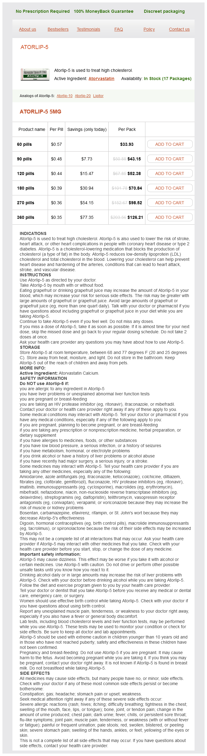
- 60 pills - $33.93
- 90 pills - $43.15
- 120 pills - $52.38
- 180 pills - $70.84
- 270 pills - $98.52
- 360 pills - $126.21
Atorlip-5 dosages: 5 mg
Atorlip-5 packs: 60 pills, 90 pills, 120 pills, 180 pills, 270 pills, 360 pills
In stock: 962
Only $0.37 per item
Description
It splits to enclose the trapezius cholesterol ratio is 2.5 purchase 5 mg atorlip-5 mastercard, the omohyoid, sternocleidomastoid, the strap muscles and the parotid gland. The superior attachment is to the external occipital protuberance, the superior nuchal lines, the mastoid tip and the zygomatic arch. The splitting of this fascial layer around the parotid forms a deep layer, which fuses with the fascia around the internal carotid artery. The boundaries are the posterior border of the sternocleidomastoid, the anterior border of the trapezius and the superior border of the inferior belly of the omohyoid muscle. It contains the cervical plexus, fibrofatty tissue, lymph nodes and the accessory nerve. It contains 80 percent of the lymph nodes of the neck, the carotid arteries, the internal jugular vein, as well as the vagus nerve. The boundaries are the lower border of the inferior belly of omohyoid, the clavicle and the posterior border sternocleidomastoid. The contents are fibrofatty tissue, the scalene muscles, the brachial plexus and the subclavian vessels, including the thyrocervical trunk. The trapezius muscle covers the cervico-occipital region and represents the posterior neck. This area is rarely involved in head and neck surgery so will not be considered in detail. Movement of the hyoid and strap muscles during swallowing elevates the fascia so that thyroid lumps characteristically move on deglutition. The deep fascia has three layers, a superficial layer, middle or visceral layer and a deep layer. This arises from the ligamentum nuchae and the spinous processes of the cervical vertebrae. It splits to enclose the postvertebral muscles, passes laterally around the scalene muscles and then forms a layer over the vertebrae. It forms the floor of the posterior triangles and allows the pharynx to glide during deglutition. Superficial cervical fascia this is a thin layer that invests the platysma muscle. Submental space this is a midline space that lies between the anterior bellies of the digastric muscles. Submandibular space the superficial boundary is the submandibular gland and digastric muscle, the deep boundary is the mylohyoid muscle. It communicates with the floor of mouth around the posterior border of the mylohyoid. It communicates through the fibres of the superior constrictor with retropharyngeal and parapharyngeal spaces. It is bounded medially by the superior constrictor and laterally by the pterygoid muscles, the mandible and deep lobe of the parotid gland. The parapharyngeal space is divided by the styloid process and its attachments into the prestyloid and poststyloid spaces.
Soapbark (Quillaia). Atorlip-5.
- Are there any interactions with medications?
- Dosing considerations for Quillaia.
- What is Quillaia?
- Are there safety concerns?
- How does Quillaia work?
Source: http://www.rxlist.com/script/main/art.asp?articlekey=96399
The mouth extends from the lips and cheeks externally to the anterior pillars of the fauces (palatoglossal arches) internally high cholesterol levels nz 5 mg atorlip-5 order overnight delivery, where it continues into the oropharynx. The floor of the mouth is formed by the mylohyoid muscles and is occupied mainly by the tongue. Opening into the mouth are three pairs of major salivary glands (parotid, submandibular and sublingual) and numerous minor salivary glands (labial, buccal, palatal, lingual). Hard palate the skeleton of the hard palate is formed by the palatine processes of the maxillae and the horizontal plates of the palatine bones. The periphery of the hard palate surrounding the necks of the teeth is termed the gingiva and a zone similarly lacking submucosa runs anteroposteriorly in the midline as a narrow, low ridge, the palatine raphe. Radiating outwards from the palatine raphe in the anterior half of the hard palate are irregular transverse ridges or rugae. The submucosa in the posterior half of the hard palate contains minor mucous salivary glands. These secrete through numerous small ducts although, bilaterally, larger ducts collecting from many of these glands often open at the paired palatine foveae. These are two sagittally elongated depressions, sometimes a few millimetres deep, which flank the midline raphe at the posterior border of the hard palate. The upper nasal surface of the hard palate is the floor of the nasal cavity and is covered by ciliated respiratory epithelium. The soft palate contains an aponeurosis, muscular tissue, vessels, nerves, lymphoid tissue and mucous glands, while some taste buds are situated on its oral aspect. A thin, fibrous palatine aponeurosis is attached to the posterior border of the hard palate. It represents the expanded tendons of the tensor veli palatini muscles and provides the fibrous skeleton of the soft palate that supports the palatine musculature. The aponeurosis is thick in the anterior two-thirds of the soft palate but very thin further back. The parotid duct drains in the region of a small parotid papilla opposite the maxillary second molar tooth. A whitish line (the linea alba) may be seen at a position related to the occlusal plane of the teeth, this hyperkeratinized line is presumably the result of continuous mild trauma during biting. Behind the molar teeth, a fold of mucosa can be seen extending from the upper to the lower alveolus, especially when the mouth is opened widely. This fold covers the pterygomandibular raphe that extends from the pterygoid hamulus to the back of the mylohyoid line. The boundary between the hard and soft palate may be distinguished by a change in colour, the soft palate being a darker red with a yellowish tint.
Specifications/Details
It is thought that a number of factors contribute to closure of the lower oesophagus including intrinsic muscle activity what type cholesterol in eggs cheap 5 mg atorlip-5 fast delivery, the encircling fibres of the right crus of the diaphragm and oblique fibres of the stomach, the thoracoabdominal pressure gradient and the angle of the oesophagogastric junction. There is an external adventitia of dense connective tissue overlying the oesophagus. A condensation of this, the phreno-oesophageal ligament, attaches the oesophagus to the diaphragmatic opening. Relationships of the oesophagus the cervical oesophagus lies posterior to the trachea and anterior to the prevertebral fascia. The recurrent laryngeal nerves ascend on each side in the tracheo-oesophageal groove. In the superior mediastinum, the oesophagus lies slightly to the left of the midline and passes behind and to the right of the aortic arch. The left subclavian artery is immediately to the left of the oesophagus as it arises from the aortic arch. On the right is the azygos vein arching from posterior to anterior over the lung root to enter the superior vena cava. The mediastinal pleura is in contact with the oesophagus, separated on the right by the azygos vein and on the left by the aortic arch and left subclavian artery. At the level of the tracheal bifurcation the oesophagus is crossed anteriorly by the left main bronchus and immediately below by the right pulmonary artery. The fibrous pericardium overlying the left atrium comes into contact with the anterior surface of the oesophagus further down. The inferior tracheobronchial nodes lie between the bifurcation of the trachea and the oesophagus. The thoracic duct enters the thorax through the right side of the aortic opening in the diaphragm and runs up behind the right margin of the oesophagus until it crosses obliquely to lie behind the left side. The two hemiazygos veins lie between the oesophagus and the prevertebral fascia and then pass to join the azygos vein on the right. Inferiorly, near the diaphragm, the aorta passes behind the oesophagus as the latter curves toward the left and turns forwards to pass through the diaphragm. The left and right vagus nerves, having branched to form the cardiac and pulmonary plexuses, come together as the oesophageal plexus and form multiple nerve trunks that descend with the oesophagus through the diaphragm. The left vagal fibres usually lie on the anterior surface and those on the right posteriorly. After the oesophagus emerges from the right crus of the diaphragm slightly to the left of the midline, it lies in the oesophageal groove on the posterior surface of the left lobe of the liver. Nerve supply to the oesophagus the striated muscle in the upper third of the oesophagus is supplied by the recurrent laryngeal nerve. The smooth muscle is supplied by parasympathetic fibres from the oesophageal branches of the vagus and recurrent laryngeal nerves. Sympathetic fibres are derived from the sympathetic trunk and come either directly from the cardiac plexus or around the blood vessels supplying the oesophagus. Pain is poorly localized and probably caused by muscle spasm rather than direct stimuli.
Syndromes
- Sucking reflex (sucks when area around mouth is touched).
- Low blood pressure
- CT or MRI scan of the neck and chest
- DHEA blood test
- Warming and cooling the inner ear with water (caloric stimulation) or air to test eye movements
- What other options you have regarding maintaining your pregnancy
- Hydronephrosis (fluid-filled kidney) can cause a smooth, spongy-feeling mass in one or both sides or toward the back (flank area).
- Heart attack
- Lumpectomy is when only the breast cancer and tissue around the cancer are removed. This is also called breast conservation therapy or partial mastectomy. Part of your breast will be left.
- Taking pain medicines
Related Products
Usage: gtt.
Additional information:
9 of 10
Votes: 312 votes
Total customer reviews: 312
Customer Reviews
Bengerd, 54 years: The systemic management of long-standing xerostomia, ¨ as in Sjogren syndrome, is summarized in Table 147. A channel is needed for each transducer pressure reading, and these are displayed in graph form with amplitude (x axis) against time (y axis). There is good theoretical evidence to support their use and in vitro experiments but no clear benefit has been demonstrated. It represents the expanded tendons of the tensor veli palatini muscles and provides the fibrous skeleton of the soft palate that supports the palatine musculature.
Myxir, 59 years: Acute suppurative sialadenitis usually affects adults, although children may rarely be affected. The external branch provides motor supply to the cricothyroid muscle, while the internal branch pierces the thyrohyoid membrane above the entrance of the superior laryngeal artery and divides into two main sensory and secretomotor branches. Multiple techniques exist Endotracheal intubation is again the most common cause for tracheal stenosis. In debilitated patients botulinum toxin injections can be performed every four to six months.
Ur-Gosh, 27 years: Lack of saliva predisposes to dental caries, gingival disease, candidosis and acute infective sialadenitis, although in children rampant dental caries may be the only initial sign of underlying salivary agenesis. The vibration of the vocal folds (phonation) constitutes the raw glottal sound source. For suction, the most widely used devices are the CarlsonCrittenden or Lashley cups, the centre of which are placed over the parotid papilla. Clinical examination of the neck in this situation has a false positive rate of between 20 and 30 percent1, 2 and a falsenegative rate of 3040 percent.
Faesul, 61 years: Videofluoroscopic evaluation in the assessment of swallowing disorders in paediatric and Deficiencies in current knowledge and areas for future research There needs to be further development of standard procedures and good intrajudge and interjudge reliability for functional investigations of dysphagia. Its medial margin is the middle layer of the fascia, laterally is the superficial layer and posterior is the anterior part of the carotid sheath. The white cell count is important and significant elevation is likely to occur in patients with impending airway obstruction. It is a challenging procedure with blanching of the vocal fold mucosa if too superficial.
Ilja, 25 years: Objective voice measurements (see Chapter 166, Objective evaluation of the voice). In a normal voice, the glottal signal is essentially periodic and composed of a fundamental frequency and its harmonics. It is often a nebulous clinical diagnosis to make because the symptoms are variable within and between subjects and objective clinical findings are by definition absent. The lesion of primary syphilis is at the site of initial inoculation and the organism can penetrate both normal and mucosal abrasions.
Flint, 32 years: Patients have a right to expect that information about them will be held in confidence by health professionals who care for them. The recurrent laryngeal nerves ascend on each side in the tracheo-oesophageal groove. When it fails, there will normally be simultaneous failure (90 percent) at the primary and/or distant sites. However, the claimant usually alleges that they failed to receive the appointment in the mail.
Contact
0673406227
dppsmyanmar@gmail.com