Actonel
Actonel 35mg
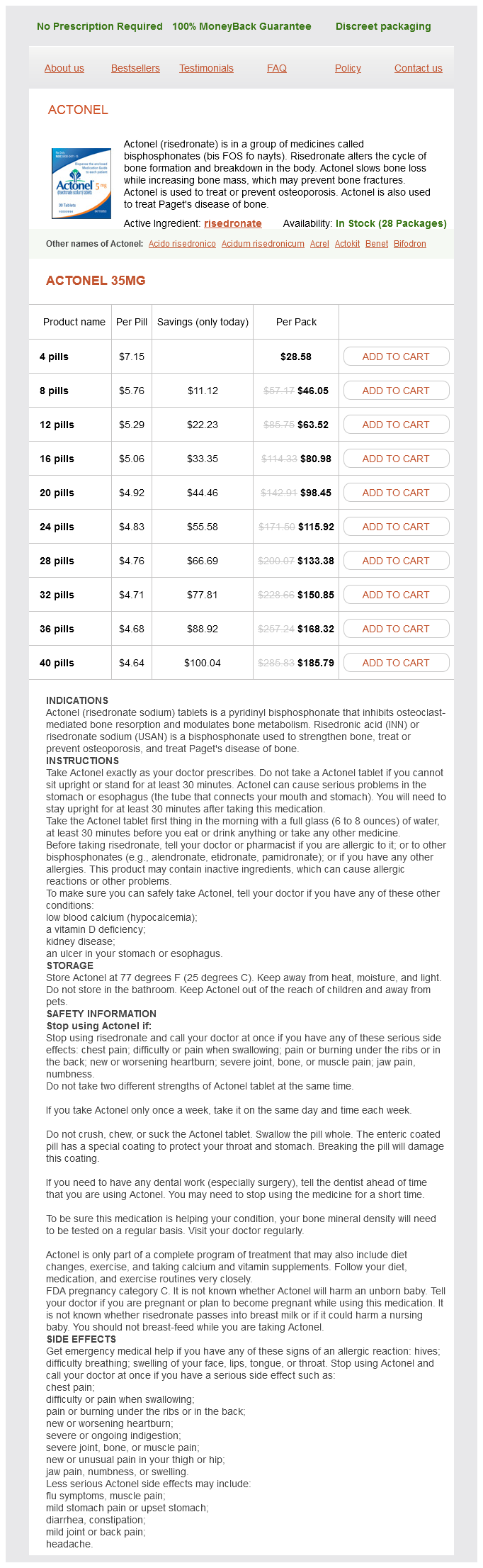
- 4 pills - $28.58
- 8 pills - $46.05
- 12 pills - $63.52
- 16 pills - $80.98
- 20 pills - $98.45
- 24 pills - $115.92
- 28 pills - $133.38
- 32 pills - $150.85
- 36 pills - $168.32
- 40 pills - $185.79
Actonel dosages: 35 mg
Actonel packs: 4 pills, 8 pills, 12 pills, 16 pills, 20 pills, 24 pills, 28 pills, 32 pills, 36 pills, 40 pills
In stock: 967
Only $4.93 per item
Description
The interpretation of cephalometric radiographs remains useful in the analysis of facial heights and maxillary medicine quiz discount 35 mg actonel otc, mandibular, and chin positions and their relationships to one another, the cranial base, and the dentition. Unfortunately, the number of anatomic landmarks that can be identified accurately in the cranio-orbitozygomatic region is limited because of the overlap of structures, which makes predictably locating these anatomic landmarks more difficult. These methods of assessment rely on the measurement of linear distances, angles, and proportions based on accurate, reliable, and reproducible anatomic landmarks found to be useful for patient evaluation. Schilli to disagreed with Cushing and enthusiastically supported the concept of surgical intervention to improve the outlook for these children. He believed that the linear "strip" craniectomy of the fused sutures would "release" the skull and allow the cranium to reshape itself and continue to grow in a normal and symmetrical fashion. With the realization that this goal was not biologically feasible, attempts were made to remove portions of the cranial vault surgically and to either leave large open areas or use the removed segments as free grafts to refashion the cranial vault shape. Problems with these methods included uncontrolled postoperative skull molding, re-ossification in to dysmorphic configurations, and the occurrence of large residual skull defects. The concept of simultaneous release of the fused suture combined with more meticulous cranial vault reshaping carried out in infants was initially suggested by Rougerie and coworkers53 and then refined by Hoffman and Mohr in 197654 for children born with unilateral coronal synostosis. Whitaker and colleagues55 proposed a more formal anterior cranial vault and orbital reshaping procedure for unilateral coronal synostosis in 1977. Marchac and associates41 published their experience with a "floating forehead" technique for simultaneous unilateral coronal and bilateral coronal suture release and forehead and upper orbital osteotomies with advancement to manage craniosynostosis in infancy. Unfortunately, the "floating forehead" technique resulted in unpredictable re-ossification of the open cranial vault areas. In addition, bitemporal constrictions or bulging and vertex of the skull concavities or bulging were frequent developments as reossification occurred. He mobilized the midface by a variety of osteotomies preformed through skin excisions directly over each osteotomy site. There is no evidence that Gillies and Harrison used any bone grafts in the surgical gaps created. This work was first presented in France at a meeting in Montpelier in 1966 and the following year at the International Plastic Surgery Meeting in Rome. He also applied an external fixation device to help maintain bony stability until healing had occurred. In 1979, Van der Meulen69 described the "medial fasciotomy" for the correction of midline facial clefting. Van der Meulen split the monobloc osteotomy vertically in the midline, removed central nasal and ethmoid bone, and then moved the two halves of the facial skeleton together for correction of the orbital hypertelorism. The widespread use of autogenous cranial bone grafting has virtually eliminated the need for rib and hip grafts when bone replacement is required in cranio-orbitozygomatic procedures. In 1968, Luhr72 introduced the use of small metal (vitalium) plates and screws to stabilize maxillofacial fractures and then osteotomies. In current practice, the use of internal miniplate and microplate and screw (titanium) fixation (of various sizes) is the preferred form of fixation when stability and three-dimensional craniomaxillofacial reconstruction of multiple osteotomized bone segments and grafts are required.
Carapa (Andiroba). Actonel.
- Fevers, herpes, intestinal worms, coughs, skin conditions, sores, ulcers, removing ticks from the head, skin parasites, arthritis, muscle and joint aches and injuries, and other conditions.
- How does Andiroba work?
- Dosing considerations for Andiroba.
- Are there safety concerns?
- What is Andiroba?
Source: http://www.rxlist.com/script/main/art.asp?articlekey=96606
Review of the blepharoplasty incisions as a surgical approach to zygomatic orbital fractures medications zofran actonel 35 mg order on-line. Computerassisted planning, stereolithographic modeling, and intraoperative navigation for complex orbital reconstruction: a descriptive study in a preliminary cohort. Thankfully, fractures in this region are infrequent, occurring among only 2% to 15% of patients with facial fractures,14 because when these fractures do occur, they can cause devastating complications because of their proximity to the brain, eyes, and nose. Complications include blindness or other forms of visual disturbance, orbital cellulitis or abscess, meningitis, brain abscess, and facial deformation. Although reports of the surgical management of the diseased frontal sinus have existed for more than 100 years,5 no consensus has yet been reached on ideal patient care after traumatic injury. The published frequency of fractures of the anterior wall, the posterior wall, and the floor of the frontal sinus varies rather widely: 43% to 61% of reported patients had anterior table fractures only; 19% to 51% had anterior and posterior table fractures; 2. The sinus may develop from one or several different sites: as a rudiment of the ethmoid air cells, as a mucosal pocket in or near the frontal recess, as an evagination of the frontal recess, or from the superior middle meatus. Secondary pneumatization begins between the ages of 6 months to 2 years and develops laterally and vertically. The sinus is radiographically identifiable by the time the child reaches the age of 6 years. Two frontal tuberosities are noted lateral to the midline and superior to the supraorbital run. The thickest area of the bone is the supraorbital rim, running from the frontozygomatic process to the nasal bones. The ethmoid plate is bound on three surfaces along the floor of the frontal bone in the midline. As the floor of the frontal bone extends laterally, it becomes concave and forms the orbital roof. The supraorbital and frontal foramina are located at the most superior portion of the orbital rim. The supratrochlear foramen is located medial to the supraorbital foramen or notch and lateral to the nasal bones. The duct of the frontal sinus empties in to the ethmoid air cells of the middle meatus of the nose. The internal concave surface of the frontal bone forms the anterior cranial fossa that houses the brain. The floor of the Physiology of the Sinus the entire surface area of the frontal sinus is covered with respiratory epithelium ranging in thickness from 0.
Specifications/Details
If the patient is able to breathe around the tube changer as it remains in the trachea treatment zenkers diverticulum 35 mg actonel purchase visa, extubation can be completed. If the patient is not able to breathe around the tube changer, the endotracheal tube is re-inserted over the tube changer in to the trachea. The endotracheal tube cuff is re-inflated, the tube changer is withdrawn, and oxygen is reconnected. After extubation, the patient is closely monitored clinically and with pulse oximetry. Arterial blood gases may be drawn 1 hour after extubation in order to verify adequate oxygenation and ventilation. Occasionally, the infecting flora, especially in a particularly severe infection with a prolonged course, will change during the course of treatment. This may be due to the selection pressure exerted by intensive antibiotic therapy or to the subsequent introduction of hospital-acquired pathogens, resulting in a nosocomial infection. Therefore, in prolonged treatments and in especially severe cases, it may be prudent to reculture infected sites so that any new or previously undetected pathogens can be identified. A draining sinus tract on to the face resulting from an untreated periapical abscess. Foreign bodies may be represented by bone plates and screws or dental or cosmetic facial implants. Epithelium may cause chronic drainage simply because an epithelialized fistulous tract has not been completely excised or because an epithelium-lined cyst has drained externally. Tumors (especially malignant ones) that become infected do not heal, which may result in chronic drainage. Distal obstruction classically refers to intestinal obstructions, but the concept can still be applied to the salivary ducts and to the natural sinus drainage pathways, such as the ostium of the maxillary sinus. If a thorough search for previously undetected pathogens turns up negative or if another cause for treatment failure cannot be found, the surgeon should consider the possibility of antibiotic failure, such as microbial resistance to empirical antibiotic therapy or the use of an incorrect dosage or route of administration for the antibiotic. Because of the necessary time delay in obtaining culture and sensitivity reports, it is occasionally necessary to change from one empirical antibiotic to another. Ideally, the surgeon should consider another of the empirical antibiotics of choice listed in Table 38-8. The input of an infectious disease consultant may also be valuable in this situation. Examples include sinusitis due to oroantral fistula or bone augmentation procedures in the posterior maxilla. The clinical diagnosis of acute sinusitis includes pain overlying the involved sinus(es), which may range from a dull ache to throbbing pain, usually constant but exacerbated by bending over or straining. Early on, the only presenting pain may involve the posterior maxillary dentition on the affected side, which may be tender to percussion. In acute sinusitis, there is profuse nasal discharge, which may range from clear to mucopurulent; in chronic sinusitis, the discharge is generally purulent and associated with nasal congestion.
Syndromes
- Eat a healthy, well-balanced diet.
- Primary care doctors
- Artificial insemination with infected semen
- Strep throat
- Surgical procedures on your palate
- Maintain a relationship with your health care provider in case of illness
- Amylase
- Carbohydrates should make up less than half of the calories you eat.
- Complete blood count with differential
- The time it was swallowed
Related Products
Usage: gtt.
Additional information:
8 of 10
Votes: 266 votes
Total customer reviews: 266
Customer Reviews
Jorn, 48 years: With persistent epiphora after trauma or surgical intervention, it is important to establish the precise point of mechanical obstruction that exists within the lacrimal drainage system. However, the two modalities that have been the most thoroughly evaluated and that have undergone the test of time are surgery and radiation therapy. For this reason, most orbital wall fractures will improve over several days of observation as the pieces self-reduce to their anatomic positions.
Sugut, 38 years: On histologic examination, sinusoidal blood spaces devoid of endothelial lining may contain a large number of multinucleated giant cells, extravasated blood cells, and hemosiderin Congenital masses (some representing cystic lesions) are classically divided based upon their location in the neck as midline or lateral and include a wide variety of specific cysts and tumors (Table 37-8). In so doing, salient clinical and radiographic features are discussed, as are the pathogenetic mechanisms supporting proliferation of some of the more aggressive odontogenic cysts and tumors. The sternocleidomastoid muscle as a muscular or myocutaneous flap for oral and facial reconstruction.
Grompel, 54 years: Often, the range of motion is decreased as the increased size of the condylar head prevents normal translation. After the incision of the skin and subcutaneous fat, the external oblique muscle is incised allowing for the visualization of the internal oblique muscle. The Gunning splint has been used to establish intermaxillary fixation for edentulous patients; this splint is essentially a denture baseplate fabricated to the existing edentulous or partially edentulous ridge; to this baseplace are attached arch bars or suspension brackets.
Contact
0673406227
dppsmyanmar@gmail.com